
Dr Marco Palombo
(e/fe)
- Ar gael fel goruchwyliwr ôl-raddedig
Timau a rolau for Marco Palombo
Uwch Ddarlithydd (Athro Cysylltiol), Pennaeth Delweddu Microstrwythurol yn CUBRIC
Uwch Ddarlithydd (Athro Cysylltiol), Sylfaenydd a Chyd-arweinydd Grŵp Cyfrifiadura Delweddau Meddygol
Trosolwyg
Bywgraffiad
Rwy'n Gymrawd Arweinwyr y Dyfodol UKRI ac yn Athro Cysylltiol (Uwch Ddarlithydd) mewn Delweddu Microstrwythurol ym Mhrifysgol Caerdydd, gyda phenodiad ar y cyd rhwng Canolfan Delweddu Ymchwil yr Ymennydd Prifysgol Caerdydd (CUBRIC) yn yr Ysgol Seicoleg, lle rwy'n Bennaeth Delweddu Microstrwythurol ac yn cyd-arwain y grŵp MicroTeam, a'r Ysgol Cyfrifiadureg a Gwybodeg, lle sefydlais a chyd-arwain y grŵp Cyfrifiadura Delweddau Meddygol.
Rwyf hefyd yn academydd arweiniol yn Hwb Oncoleg Manwl Rhinddisgyblaethol Caerdydd (IPOCH) ac rwyf wedi bod yn aelod o'r Ganolfan Deallusrwydd Artiffisial, Roboteg a Systemau Dynol-Peiriant (IROHMS) ym Mhrifysgol Caerdydd lle roeddwn i'n cyd-gadeirio'r Gweithgor "Human-centric AI for Medical Imaging".
Fi yw Prif Olygydd Sefydlu Medical Sensors and Imaging, cyfnodolyn mynediad agored IOP Press mewn cydweithrediad â'r Sefydliad Ffiseg a Pheirianneg mewn Meddygaeth (IPEM) gyda'r nod o hyrwyddo a lledaenu ymchwil o ansawdd uchel ar dechnolegau synhwyro a delweddu arloesol sy'n gwella diagnosis meddygol, monitro a gofal cleifion.
Mae gen i B.Sc., M.Sc. a PhD mewn Ffiseg, gydag arbenigedd mewn modelu bioffisegol, dysgu peiriannau, modelu cyfrifiadurol, delweddu meddygol, a dadansoddi data.
Ymchwil
Rwy'n arwain y rhaglen ymchwil Delweddu Microstrwythur yn CUBRIC. Fy niddordeb ymchwil yw cyfuno Ffiseg, Cyfrifiadureg a Niwrowyddoniaeth i ddatblygu technolegau delweddu anfewnwthiol ar gyfer diagnosis cynnar a prognosis o gyflyrau niwrolegol a seiciatrig.
Tuag at y nod hwn, mae fy nhîm (gweler tab Ymchwil) a minnau'n cyfuno modelu cyfrifiadurol, MRI a dysgu peiriannau modern i arloesi arloesiadau allweddol mewn delweddu microstrwythur a histoleg anfewnwthiol yr ymennydd (gyda ffocws penodol ar fater llwyd):
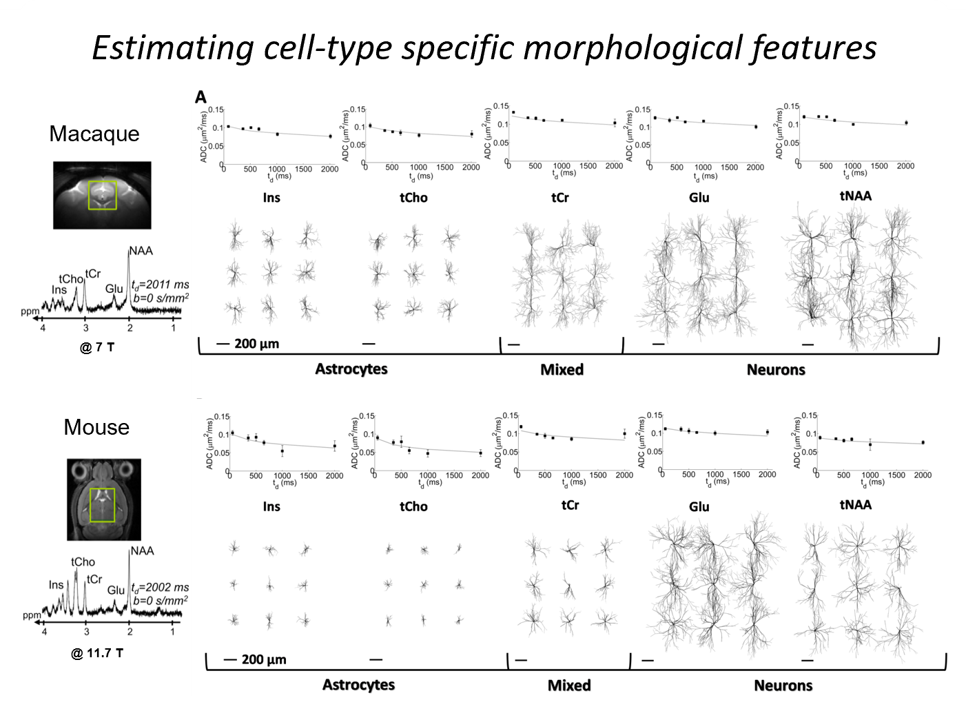
Yr arddangosiad cyntaf o feintioli anfewnwthiol morffoleg celloedd ymennydd cymhleth, gan ddefnyddio sbectrosgopeg MR wedi'i bwysoli trylediad a modelu cyfrifiadurol (Palombo et al., PNAS 2016);
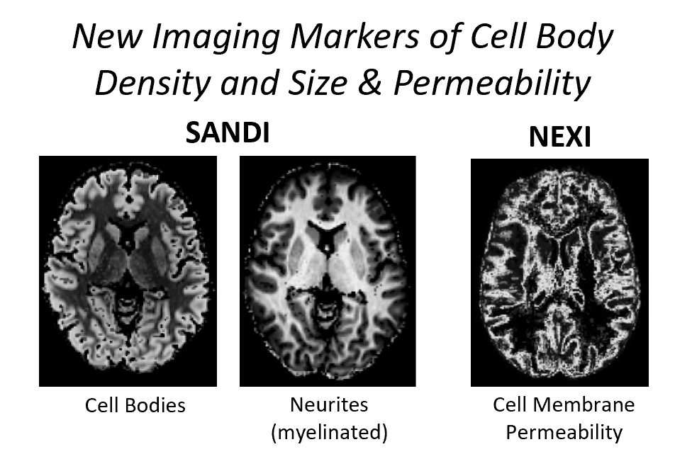
Mapio maint a dwysedd corff celloedd, gan ddefnyddio SANDI (Palombo et al. Neuroimage 2020, Ianus et al. Neuroimage 2022); mapio cyfnewid dŵr, gan ddefnyddio NEXI (Jelescu et al. Neuroimage 2022; Uhl et al. Delweddu Niwrowyddoniaeth 2024);
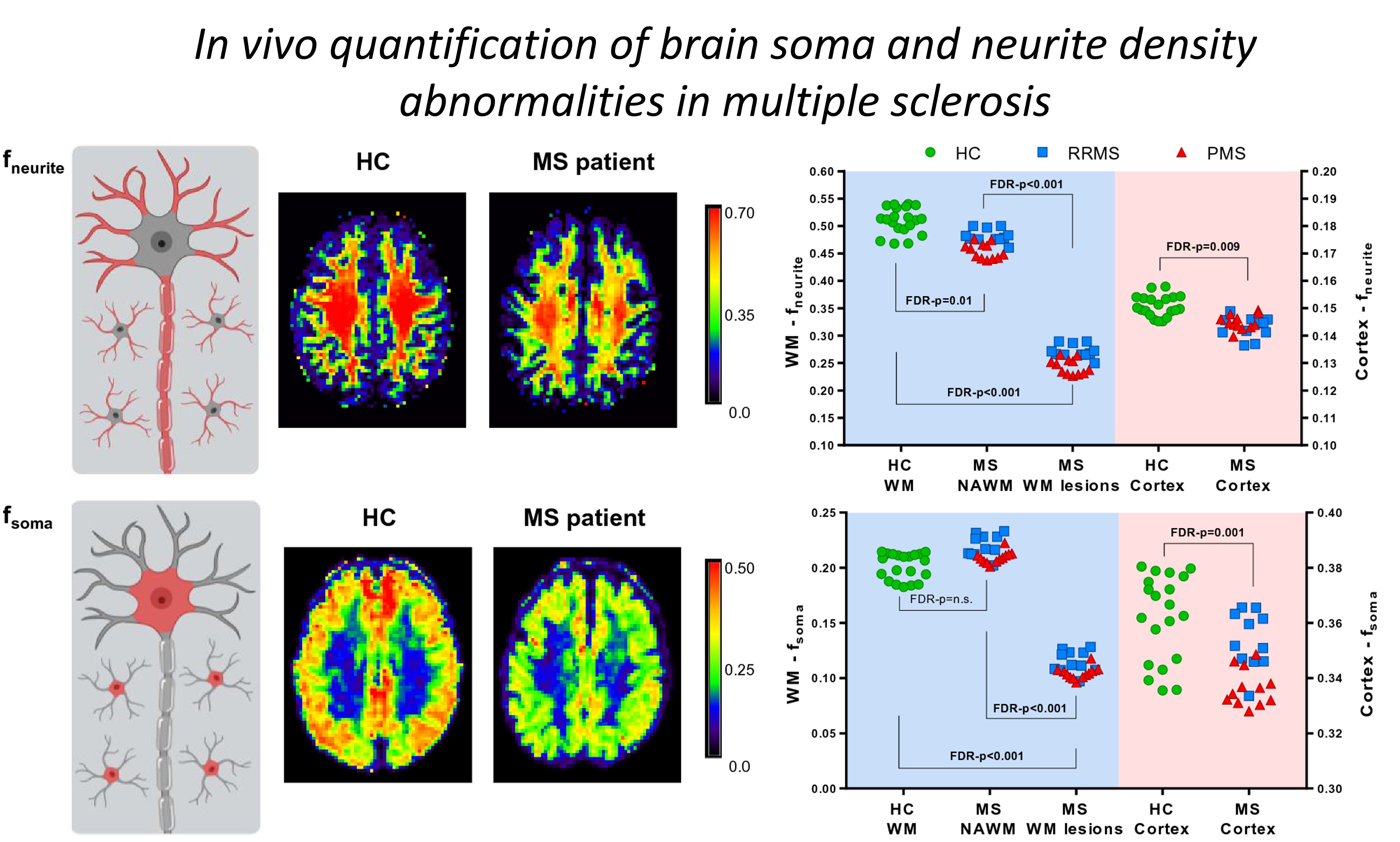
Cyfieithu delweddu maint a dwysedd corff celloedd gan ddefnyddio SANDI ar sganwyr 3T clinigol i nodweddu patholeg Sglerosis Ymledol (MS) (Schiavi et al. Human Brain Mapping 2023, Magoni et al. Journal of Neurology 2023, Barakovic et al. Nature Sci Rep 2024);
Effeithiau cyfyngu a chyfnewid delweddu gan ddefnyddio cymhareb amserol trylediad, TDR (Warner et al. Neuroimage 2023) a dibyniaeth amser trylediad dŵr a metabolit cyfunol (Mougel et al. Imaging Neuroscience 2024);
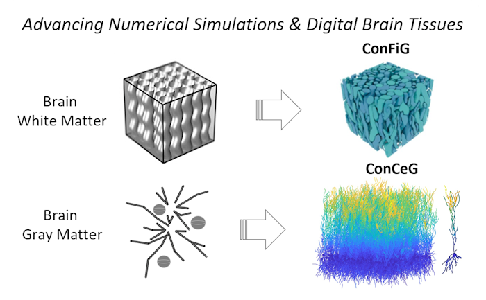
Modelau cynhyrchiol cyntaf o'i fath o morffolegau celloedd yr ymennydd cymhleth (Palombo et al., Neuroimage 2019), bwndeli axonal gyda ConFiG (Callaghan et al. Neuroimage 2020) a mater llwyd yr ymennydd gyda ConCeG (Aird-Rossiter et al. Bioleg Cyfathrebu 2025; Aird-Rossiter et al. arXiv 2026) ar gyfer cynhyrchu modelau cyfrifiadurol ultra-realistig o ficrostrwythur yr ymennydd, sy'n hanfodol ar gyfer efelychiadau rhifiadol mwy realistig (e.e. Monte Carlo)
Mae gwaith diweddar yn canolbwyntio ar gyfuno modelau cyfrifiadurol o'r fath, efelychiadau Monte Carlo a dysgu peiriannau ar gyfer delweddu microstrwythur y genhedlaeth nesaf, e.e.:
Mapiau o athreiddedd axonal fel marciwr delweddu newydd o demyelination (Hill et al. Neuroimage 2021);
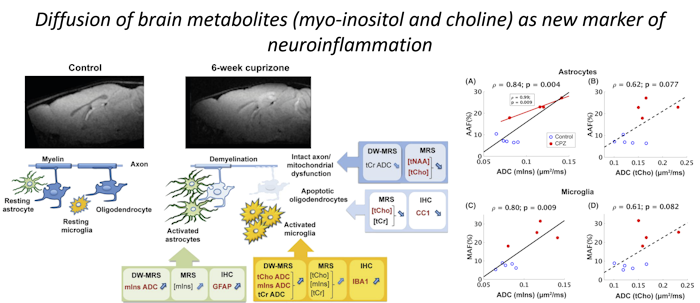
Meintioli in vivo o adweithedd glial a niwrolid (Ligneul et al., Neuroimage 2019, Genovese et al., NMR Biomed 2021);
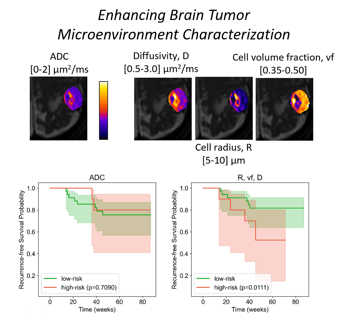
Delweddu microstrwythur canser-benodol i asesu ymateb tiwmor yr ymennydd i therapi radio / proton (Buizza et al., Ffiseg Feddygol 2020, Morelli et al. Ffiseg Feddygol 2023); canser endometriaidd (Maiuro et al. Ffiseg Feddygol 2025); ac efelychiadau a fframweithiau delweddu microstrwythur wedi'u teilwra ar gyfer canser yr afu (Grussu et al. Meddygaeth Cyfathrebu 2025, Grigoriu et al. Bioleg Cyfathrebu 2025, Voronova et al. Dadansoddiad Delwedd Feddygol 2025)
Amcangyfrif ar y cyd o briodweddau trylediad ac ymlacio canser y prostad gyda rVERDICT (Palombo et al. Nature Sci. Rep. 2023), a nodweddu microstrwythurol uwch o ganser y prostad gan ddefnyddio graddiannau cryf iawn (Molendowska et al. NMR Biomed 2024)

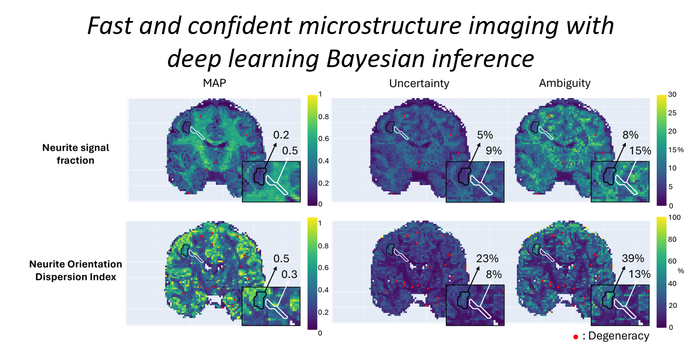
Casgliad Bayesian effeithlon gan ddefnyddio dysgu dwfn ar gyfer meintioli ansicrwydd a dirywiad mewn delweddu microstrwythur gan ddefnyddio μGUIDE (Jallais a Palombo, eLife 2024)
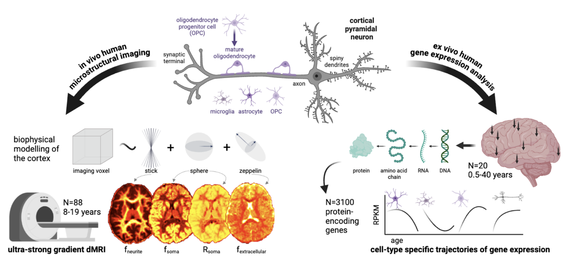
Nodweddu uwch microstrwythur yr ymennydd yn ystod niwrodatblygiad iach (Genc et al. Cyfathrebu Natur 2025, Ligneul et al. eLife 2025, Karat et al. Bioleg Cyfathrebu 2026), plastigrwydd (Griffa et al. Bioleg PLoS 2026) a niwroddirywiad, fel Clefyd Huntington (Ioakeimidis et al. eLife 2026)
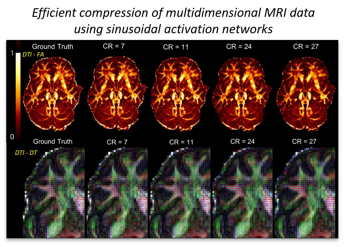
Cywasgu data effeithlon o ddata MRI aml-ddimensiwn gyda SirenMRI (Mancini et al, Nodiadau Darlith mewn Cyfrifiadureg 2022)
Cod ffynhonnell agored:
- Generadur Cell Rhithwir: https://github.com/palombom/Virtual-Cell-Generator-v1.0
- SANDI: https://github.com/palombom/SANDI-Matlab-Toolbox-Latest-Release
- NEXI: https://github.com/QuentinUhl/nexi
- Ymlacio-DYFARNIAD: https://github.com/palombom/Relaxation-VERDICT-Matlab-Toolbox-v1.0
- Aml-TE SANDI: https://github.com/palombom/MultiTE-SANDI-Matlab-Toolbox-v1.0
- CANLLAW: https://github.com/mjallais/uGUIDE
- SirenMRI: https://github.com/palombom/SirenMRI
Cyllid
Mae ein hymchwil yn cael ei gefnogi gan ystod o gyrff cyllido a phartneriaid diwydiannol:
Ymchwil ac Arloesi y DU
- 2025-2028: Adnewyddu Cymrodoriaeth Arweinwyr y Dyfodol UKRI: MR/T020296/2, (Prif Ymchwilydd Palombo), ~£700k
- 2022-2025: UKRI MRC: MR/W031566/1, (Cyd-Brif Ymchwilydd Palombo), ~£1m
- 2020-2025: Cymrodoriaeth Arweinwyr y Dyfodol UKRI: MR/T020296/1 & 2, (Prif Ymchwilydd Palombo), ~£1.3m
- 2022-2024: UKRI BBSRC: BB/X005089/1, (Prif Ymchwilydd Palombo), ~£22k
- 2022-2027: Ysgoloriaethau DTP EPSRC UKRI (Lewis Kitchingman a Jiří Benáček), ~£130k
Ymddiriedolaeth Wellcome:
- 2025-2033: Gwobr Darganfod Wellcome [317797/Z/24/Z] "Democrateiddio Ymchwil Niwrodelweddu gydag MRI" (Cyd-Brif Ymchwilydd Palombo), ~£4m
Ymchwil Canser Cymru ac Ymddiriedolaeth GIG Prifysgol Felindre:
- Astudiaeth MIMOSA 2024- 2027 (Cyd-ymgeisydd Arweiniol Palombo), ~£350k
Partneriaethau Strategol gyda Diwydiant
- GlaxoSmithKline Plc (GSK) - Ysgoloriaeth PhD (Elise Gwyther)
- F. Hoffmann-La Roche Ltd (Roche) - Prosiect ymchwil DEPICT (2024-2027)
- Siemens Healthineers Cyf
Cyngor Ymchwil Awstralia (ARC)
- Prosiect Darganfod 2025- 2028 "Modelau mathemategol newydd ar gyfer delweddu microstrwythur meinwe'r ymennydd" (Cyd-Brif Ymchwilydd Palombo), ~£300k
Cyhoeddiad
2026
- Şimşek, K. et al. 2026. The role of dendritic spines in water exchange measurements with diffusion MRI: time‐dependent single diffusion encoding MRI. NMR in Biomedicine 39 (10) e70382. (10.1002/nbm.70382)
- Hiscox, L. et al. 2026. 88 Slip interface imaging in glioblastoma: a pilot study of tumour-brain adhesion and surgical resectability [Abstract]. Neuro-Oncology 28 (S1) noag172.029. (10.1093/neuonc/noag172.029)
- Sen, S. et al., 2026. microTorch: A software package for fast and flexible self-supervised diffusion MRI model fitting. Journal of Open Research Software 14 (1) 59. (10.5334/jors.736)
- Golten, J. et al. 2026. 20 Updates on Magnetic Resonance Imaging assessment of tumour MicrOStructure in GlioblastomA: A cross-sectional study (The MIMOSA Study) [Abstract]. Neuro-Oncology 28 (S1) noag172.058. (10.1093/neuonc/noag172.058)
- Yang, Z. et al., 2026. Placental blood-flow velocity quantification from diffusion MRI. Magnetic Resonance in Medicine (10.1002/mrm.70551)
- Chakwizira, A. et al., 2026. The role of dendritic spines in water exchange measurements with diffusion MRI: Double diffusion encoding and free‐waveform MRI. NMR in Biomedicine 39 (7) e70315. (10.1002/nbm.70315)
- Voronova, A. K. et al., 2026. Simulation‐informed evaluation of microvascular parameter mapping for diffusion MR imaging of solid tumours. Magnetic Resonance in Medicine 96 (1), pp.387-402. (10.1002/mrm.70318)
- Jallais, M. et al. 2026. Bayesian insights into exchange and restriction in gray matter diffusion MRI. Imaging Neuroscience 4 IMAG.a.1317. (10.1162/imag.a.1317)
- Griffa, G. et al., 2026. Learning engages transient and sustained cellular mechanisms in the human brain. PLoS Biology 24 (6) e3003861. (10.1371/journal.pbio.3003861)
- Szczepankiewicz, F. et al., 2026. Microstructure imaging of prostate cancer by diffusion MRI. Magnetic Resonance Materials in Physics, Biology and Medicine (10.1007/s10334-026-01367-2)
- Palombo, M. et al. 2026. ESR Essentials: diffusion-weighted MRI - Practice recommendations by the European Society for Magnetic Resonance in Medicine and Biology. European Radiology 36 , pp.2198-2208. (10.1007/s00330-025-12033-x)
- Karat, B. G. et al., 2026. Microstructural variation of hippocampal substructures across childhood and adolescence quantified with high-gradient diffusion MRI. Communications Biology 9 416. (10.1038/s42003-026-09622-x)
2025
- Grigoriou, A. et al., 2025. Histology-informed microstructural diffusion simulations for MRI cancer characterisation—the Histo-μSim framework. Communications Biology 8 1695. (10.1038/s42003-025-09096-3)
- Grussu, F. et al., 2025. Clinically feasible liver tumour cell size measurement through histology-informed in vivo diffusion MRI. Communications Medicine 5 535. (10.1038/s43856-025-01246-2)
- Bliakharskaia, E. et al., 2025. Exploring the contribution of gray matter microstructure to R1 contrast via multi-compartment diffusion modelling in the healthy brain. NeuroImage 320 121466. (10.1016/j.neuroimage.2025.121466)
- Ligneul, C. et al., 2025. Diffusion MRS tracks distinct trajectories of neuronal development in the cerebellum and thalamus of rat neonates. eLife 13 RP96625. (10.7554/eLife.96625.4)
- McCloskey, H. et al. 2025. Quantified head-ball impacts in soccer: a preliminary, prospective study. Neurotrauma Reports 6 (1), pp.928-943. (10.1177/2689288x251380145)
- Ioakeimidis, V. et al. 2025. In vivo mapping of striatal neurodegeneration in Huntington's disease with Soma and Neurite Density Imaging. eLife 14 RP107661. (10.7554/eLife.107661.1)
- Schilling, K. G. et al., 2025. Characterization of neurite and soma organization in the brain and spinal cord with diffusion MRI. Imaging Neuroscience 3 IMAG.a.111.. (10.1162/imag.a.111)
- Maiuro, A. et al., 2025. Endometrial cancer tissue features clusterization by kurtosis MRI. Medical Physics 52 (5), pp.2898-2908. (10.1002/mp.17718)
- Şimşek, K. et al. 2025. Age-trajectories of higher-order diffusion properties of major brain metabolites in cerebral and cerebellar grey matter using in vivo diffusion-weighted MR spectroscopy at 3T. Aging Cell 24 (5) e14477. (10.1111/acel.14477)
- Voronova, A. K. et al., 2025. SpinFlowSim: A blood flow simulation framework for histology-informed diffusion MRI microvasculature mapping in cancer. Medical Image Analysis 102 103531. (10.1016/j.media.2025.103531)
- Erin, E. et al., 2025. Improving image reconstruction for ultra-fast ptychographic acquisitions via deep learning denoising. Presented at: 15th International Conference on Synchrotron Radiation Instrumentation (SRI 2024) Hamburg, Germany 26-30 August 2024. Vol. 3010.Vol. 1. IOP Publishing. (10.1088/1742-6596/3010/1/012172)
- Maiuro, A. et al., 2025. New functional MRI experiments based on fractional diffusion representation show independent and complementary contrast to diffusion-weighted and blood-oxygen-level-dependent functional MRI. Applied Sciences 15 (9) 4930. (10.3390/app15094930)
- Cicimen, A. G. et al., 2025. Image quality transfer of diffusion MRI guided By high-resolution structural MRI. Presented at: CDMRI 2024 Marrakesh, Morocco 06 October 2024. Published in: Chamberland, M. et al., Computational Diffusion MRI. Lecture Notes in Computer Science Vol. 15171. Springer Nature Switzerland. , pp.106-118. (10.1007/978-3-031-86920-4_10)
- Genc, S. et al. 2025. MRI signatures of cortical microstructure in human development align with oligodendrocyte cell-type expression. Nature Communications 16 3317. (10.1038/s41467-025-58604-w)
- Ioakeimidis, V. et al. 2025. In vivo mapping of striatal microstructure in Huntington's disease with Soma and Neurite Density Imaging. MedRXiv (10.1101/2025.03.17.25324107)
- Jones, D. K. et al. 2025. Low field, high impact: Democratizing MRI for clinical and research innovation. BJR Open 7 (1) tzaf022. (10.1093/bjro/tzaf022)
- Preziosa, P. et al., 2025. Soma and neurite density abnormalities of paramagnetic rim lesions and core-sign lesions in multiple sclerosis. Journal of Neurology 272 145. (10.1007/s00415-025-12887-7)
- Dyrby, T. B. et al., 2025. Tractography validation Part 1: Foundations, numerical simulations, and phantom models. In: Dell'acqua, F. , Descoteaux, M. and Leemans, A. eds. Handbook of Diffusion MR Tractography. Elsevier. , pp.485-509. (10.1016/B978-0-12-818894-1.00017-3)
- Dyrby, T. B. et al., 2025. Tractography validation Part 2: The use of anatomical model systems and measures for validation. In: Dell'acqua, F. , Descoteaux, M. and Leemans, A. eds. Handbook of Diffusion MR Tractography. Elsevier. , pp.511-542. (10.1016/B978-0-12-818894-1.00020-3)
- Aird-Rossiter, C. et al. 2025. Decoding Gray Matter: large-scale analysis of brain cell morphometry to inform microstructural modeling of diffusion MR signals. Communications Biology 9 138. (10.1038/s42003-025-09353-5)
2024
- Jallais, M. and Palombo, M. 2024. Introducing µGUIDE for quantitative imaging via generalized uncertainty-driven inference using deep learning. eLife 13 RP101069. (10.7554/elife.101069.3)
- Molendowska, M. et al. 2024. Diffusion MRI in prostate cancer with ultra-strong whole body gradients. NMR in Biomedicine (10.1002/nbm.5229)
- Ioakeimidis, V. et al. 2024. Protocol for a randomised controlled unblinded feasibility trial of HD-DRUM, a rhythmic movement training application for cognitive and motor symptoms in people with Huntington’s disease. BMJ Open 14 (7) e082161. (10.1136/bmjopen-2023-082161)
- Langkammer, C. et al., 2024. ESMRMB 2024 focus topic: MR beyond trends—fact-checking MR. Magnetic Resonance Materials in Physics, Biology and Medicine 37 , pp.321-322. (10.1007/s10334-024-01177-4)
- Cipiccia, S. et al., 2024. Fast X-ray ptychography: towards nanoscale imaging of large volume of brain. European Physical Journal Plus 139 (5) 434. (10.1140/epjp/s13360-024-05224-w)
- Barakovic, M. et al., 2024. A novel imaging marker of cortical “cellularity” in multiple sclerosis patients. Scientific Reports 14 (1) 9848. (10.1038/s41598-024-60497-6)
- Preziosa, P. et al., 2024. In-vivo assessment of Cellular Soma and Neurite Density Abnormalities in Multiple Sclerosis Paramagnetic Rim and Core-sign Lesions (P11-6.006). Neurology 102 (17S1) P11-6.006. (10.1212/WNL.0000000000205130)
- Mougel, E. , Valette, J. and Palombo, M. 2024. Investigating exchange, structural disorder and restriction in Gray Matter via water and metabolites diffusivity and kurtosis time-dependence. Imaging Neuroscience 2 , pp.1-14. (10.1162/imag_a_00123)
- Ligneul, C. et al., 2024. Diffusion‐weighted MR spectroscopy: Consensus, recommendations, and resources from acquisition to modeling. Magnetic Resonance in Medicine 91 (3), pp.860-885. (10.1002/mrm.29877)
- Uhl, Q. et al., 2024. Quantifying human gray matter microstructure using neurite exchange imaging (NEXI) and 300 mT/m gradients. Imaging Neuroscience 2 , pp.1-19. (10.1162/imag_a_00104)
- Lou, J. et al. 2024. Predicting radiologists' gaze with computational saliency models in mammogram reading. IEEE Transactions on Multimedia 26 , pp.256-269. (10.1109/TMM.2023.3263553)
2023
- Endt, S. et al. 2023. In vivo myelin water quantification using diffusion–relaxation correlation MRI: A comparison of 1D and 2D methods. Applied Magnetic Resonance 54 , pp.1571-1588. (10.1007/s00723-023-01584-1)
- Ioakeimidis, V. et al. 2023. Protocol for a randomised controlled feasibility trial of HD-DRUM, a rhythmic movement training application for cognitive and motor symptoms in people with Huntington's disease. [Online].medRxiv: medRxiv. (10.1101/2023.11.15.23298581)Available at: https://doi.org/10.1101/2023.11.15.23298581.
- Schiavi, S. et al., 2023. Mapping tissue microstructure across the human brain on a clinical scanner with soma and neurite density image metrics. Human Brain Mapping 44 (13), pp.4792-4811. (10.1002/hbm.26416)
- Reddaway, J. et al. 2023. Microglial morphometric analysis: so many options, so little consistency. Frontiers in Neuroinformatics 17 1211188. (10.3389/fninf.2023.1211188)
- Örzsik, B. et al., 2023. Higher order diffusion imaging as a putative index of human sleep-related microstructural changes and glymphatic clearance. NeuroImage 274 120124. (10.1016/j.neuroimage.2023.120124)
- Morelli, L. et al., 2023. Microstructural parameters from DW-MRI for tumour characterization and local recurrence prediction in particle therapy of skull-base chordoma. Medical Physics 50 (5), pp.2900-2913. (10.1002/mp.16202)
- Figini, M. et al., 2023. Comprehensive brain tumour characterization with VERDICT-MRI: evaluation of cellular and vascular measures validated by histology. Cancers 15 (9) 2490. (10.3390/cancers15092490)
- Spindler, M. et al., 2023. Dysfunction of the hypothalamic-pituitary adrenal axis and its influence on aging: the role of the hypothalamus. Scientific Reports 13 (1) 6866. (10.1038/s41598-023-33922-5)
- Warner, W. et al., 2023. Temporal Diffusion Ratio (TDR) for imaging restricted diffusion: optimisation and pre-clinical demonstration. NeuroImage 269 119930. (10.1016/j.neuroimage.2023.119930)
- Palombo, M. et al. 2023. Joint estimation of relaxation and diffusion tissue parameters for prostate cancer with relaxation-VERDICT MRI. Scientific Reports 13 (1) 2999. (10.1038/s41598-023-30182-1)
- Margoni, M. et al., 2023. In vivo quantification of brain soma and neurite density abnormalities in multiple sclerosis. Journal of Neurology 270 (1), pp.433–445. (10.1007/s00415-022-11386-3)
2022
- Schilling, K. G. et al., 2022. Minimal number of sampling directions for robust measures of the spherical mean diffusion weighted signal: Effects of sampling directions, b-value, signal-to-noise ratio, hardware, and fitting strategy. Magnetic Resonance Imaging 94 , pp.25-35. (10.1016/j.mri.2022.07.015)
- Lim, J. P. et al., 2022. Fitting a directional microstructure model to diffusion-relaxation mri data with self-supervised machine learning. Lecture Notes in Computer Science 13722 , pp.77-88. (10.1007/978-3-031-21206-2_7)
- Jelescu, I. O. et al., 2022. Neurite Exchange Imaging (NEXI): A minimal model of diffusion in gray matter with inter-compartment water exchange. NeuroImage 256 119277.
- Ianus, A. et al., 2022. Soma and Neurite Density MRI (SANDI) of the in-vivo mouse brain and comparison with the Allen Brain Atlas. NeuroImage 254 119135. (10.1016/j.neuroimage.2022.119135)
- Gyori, N. et al., 2022. Training data distribution significantly impacts the estimation of tissue microstructure with machine learning. Magnetic Resonance in Medicine 87 (2), pp.932-947. (10.1002/mrm.29014)
- Palombo, M. et al. 2022. Transient anomalous diffusion MRI measurement discriminates porous polymeric matrices characterized by different sub-microstructures and fractal dimension. Gels 8 (2) 95. (10.3390/gels8020095)
2021
- Slator, P. et al. 2021. Combined diffusion-relaxometry microstructure imaging: Current status and future prospects. Magnetic Resonance in Medicine 86 (6), pp.2987-3011. (10.1002/mrm.28963)
- Kerkelaa, L. et al., 2021. Comparative analysis of signal models for microscopic fractional anisotropy estimation using q-space trajectory encoding. NeuroImage 242 118445. (10.1016/j.neuroimage.2021.118445)
- Ianus, A. et al., 2021. Mapping complex cell morphology in the grey matter with double diffusion encoding MR: A simulation study. NeuroImage 241 118424. (10.1016/j.neuroimage.2021.118424)
- De Luca, A. et al., 2021. On the generalizability of diffusion MRI signal representations across acquisition parameters, sequences and tissue types: chronicles of the MEMENTO challenge. NeuroImage 240 118367. (10.1016/j.neuroimage.2021.118367)
- Grussu, F. et al., 2021. Deep learning model fitting for diffusion-relaxometry: a comparative study. In: Gyori, N. et al., Computational Diffusion MRI. Mathematics and Visualization. Mathematics and Visualization Cham: Springer. , pp.159-172. (10.1007/978-3-030-73018-5_13)
- Afzali, M. et al. 2021. SPHERIOUSLY? The challenges of estimating sphere radius non-invasively in the human brain from diffusion MRI. NeuroImage 237 118183. (10.1016/j.neuroimage.2021.118183)
- Slator, P. et al. 2021. Data-driven multi-contrast spectral microstructure imaging with InSpect: INtegrated SPECTral component estimation and mapping. Medical Image Analysis 71 102045. (10.1016/j.media.2021.102045)
- Palombo, M. et al. 2021. Joint estimation of relaxation and diffusion tissue parameters for prostate cancer grading with relaxation-VERDICT MRI. [Online].medRxiv: Cold Spring Harbor Laboratory. (10.1101/2021.06.24.21259440)Available at: https://doi.org/10.1101/2021.06.24.21259440.
- Perot, J. et al., 2021. Identification of the key role of white matter alteration in the pathogenesis of Huntington’s Disease. [Online].bioRxiv: Cold Spring Harbor Laboratory. (10.1101/2021.06.21.449242)Available at: https://doi.org/10.1101/2021.06.21.449242.
- Genovese, G. et al., 2021. Inflammation-driven glial alterations in the cuprizone mouse model probed with diffusion-weighted magnetic resonance spectroscopy at 11.7 T. NMR in Biomedicine 34 (4) e4480. (10.1002/nbm.4480)
- Valindria, V. et al., 2021. Synthetic Q-Space learning with deep regression networks for prostate cancer characterisation with VERDICT. Presented at: 2021 IEEE 18th International Symposium on Biomedical Imaging Nice, France 13-16 April 2021. 2021 IEEE 18th International Symposium on Biomedical Imaging (ISBI). IEEE. , pp.50-54. (10.1109/ISBI48211.2021.9434096)
- Buizza, G. et al., 2021. Improving the characterization of meningioma microstructure in proton therapy from conventional apparent diffusion coefficient measurements using Monte Carlo simulations of diffusion MRI. Medical Physics 48 (3), pp.1250-1261. (10.1002/mp.14689)
- Callaghan, R. et al., 2021. Impact of within-voxel heterogeneity in fibre geometry on spherical deconvolution. ArXiv (10.48550/arXiv.2103.08237)
- Martins, J. P. d. A. et al., 2021. Neural networks for parameter estimation in microstructural MRI: a study with a high-dimensional diffusion-relaxation model of white matter microstructure. [Online].bioRxiv: Cold Spring Harbor Laboratory. (10.1101/2021.03.12.435163)Available at: https://doi.org/10.1101/2021.03.12.435163.
- Palombo, M. et al. 2021. Corrigendum to “SANDI: A compartment-based model for non-invasive apparent soma and neurite imaging by diffusion MRI” [Neuroimage 215 (2020), 116835]. NeuroImage 226 117612. (10.1016/j.neuroimage.2020.117612)
- Henriques, R. N. et al., 2021. Double diffusion encoding and applications for biomedical imaging. Journal of Neuroscience Methods 348 108989. (10.1016/j.jneumeth.2020.108989)
- Hill, I. et al., 2021. Machine learning based white matter models with permeability: An experimental study in cuprizone treated in-vivo mouse model of axonal demyelination.. NeuroImage 224 117425. (10.1016/j.neuroimage.2020.117425)
2020
- Ning, L. et al., 2020. Cross-scanner and cross-protocol multi-shell diffusion MRI data harmonization: algorithms and result. NeuroImage 221 117128. (10.1016/j.neuroimage.2020.117128)
- Pizzolato, M. et al., 2020. Acquiring and predicting multidimensional diffusion (MUDI) data: an open challenge. Presented at: MICCAI Workshop Shenzhen, China Oct 2019. Published in: Bonet-Carne, E. et al., Computational Diffusion MRI. Mathematics and Visualization Springer. , pp.195-208. (10.1007/978-3-030-52893-5_17)
- Callaghan, R. et al., 2020. ConFiG: Contextual Fibre Growth to generate realistic axonal packing for diffusion MRI simulation. NeuroImage 220 117107. (10.1016/j.neuroimage.2020.117107)
- Jelescu, I. O. et al., 2020. Challenges for biophysical modeling of microstructure. Journal of Neuroscience Methods 344 108861. (10.1016/j.jneumeth.2020.108861)
- Slator, P. J. et al. 2020. Data-driven multi-contrast spectral microstructure imaging with InSpect. Presented at: MICCAI: International Conference on Medical Image Computing and Computer-Assisted Intervention Lima, Peru 4–8 October, 2020. Published in: Martel, A. et al., Medical Image Computing and Computer Assisted Intervention – MICCAI 2020.. Vol. 12266.Cham: Springer. , pp.375-385. (10.1007/978-3-030-59725-2_36)
- Warner, R. W. et al., 2020. Optimisation of Temporal Diffusion Ratio (TDR) to maximise its potential to map large axons: Insight from simulations. Presented at: ISMRM and SMRT Virtual Conference and Exhibition 8-14 August 2020.
- Palombo, M. et al. 2020. SANDI: a compartment-based model for non-invasive apparent soma and neurite imaging by diffusion MRI.. NeuroImage 215 116835. (10.1016/j.neuroimage.2020.116835)
- Vincent, M. , Palombo, M. and Valette, J. 2020. Revisiting double diffusion encoding MRS in the mouse brain at 11.7T: Which microstructural features are we sensitive to?. NeuroImage 207 116399. (10.1016/j.neuroimage.2019.116399)
- Capuani, S. and Palombo, M. 2020. Mini review on anomalous diffusion by MRI: Potential advantages, pitfalls, limitations, nomenclature, and correct interpretation of literature. Frontiers in Physics 7 248. (10.3389/fphy.2019.00248)
- Guerreri, M. et al., 2020. Tortuosity assumption not the cause of NODDI’s incompatibility with tensor-valued diffusion encoding. Presented at: ISMRM and SMRT Virtual Conference and Exhibition 8-14 August 2020.
- Palombo, M. and Singh, S. 2020. Relaxed-VERDICT: decoupling relaxation and diffusion for comprehensive microstructure characterization of prostate cancer.. Presented at: ISMRM & SMRT Virtual Conference & Exhibition 2020 Online 8-14 August 2020.
2019
- Slator, P. et al. 2019. Combined diffusion-relaxometry MRI to identify dysfunction in the human placenta. Magnetic Resonance in Medicine 82 (1), pp.95-106. (10.1002/mrm.27733)
- Callaghan, R. et al., 2019. Contextual fibre growth to generate realistic axonal packing for diffusion MRI simulation. Presented at: IPMI: 26th International Conference on Information Processing in Medical Imaging Hong Kong, China 2-7-June 2019. Published in: Chung, A. et al., Information Processing in Medical Imaging Proceedings. Vol. 11492.Lecture Notes in Computer Science Springer. , pp.429-440. (10.1007/978-3-030-20351-1_33)
- Slator, P. et al. 2019. InSpect: INtegrated SPECTral component estimation and mapping for multi-contrast microstructural MRI. Presented at: IPMI 2019: International Conference on Information Processing in Medical Imaging Hong Kong 2-7 June 2019. Published in: Chung, A. C. S. et al., Information Processing in Medical Imaging: 26th International Conference, IPMI 2019, Hong Kong, China, June 2–7, 2019, Proceedings. Springer. , pp.755-766. (10.1007/978-3-030-20351-1_59)
- Ianus, A. et al., 2019. Effect of cell complexity and size on diffusion MRI signal: a simulation study. Presented at: ISMRM 27th Annual Meeting and Exhibition Montreal, QC, Canada 11-16 May 2019.
- Ning, L. et al., 2019. Muti-shell diffusion MRI harmonisation and enhancement challenge (MUSHAC): Progress and results. Presented at: MICCAI 2018 Granada, Spain 16-20 September 2018. Published in: Bonet-Carne, E. et al., Computational Diffusion MRI. Vol. 1.Mathematics and Visualization Cham: Springer. , pp.217-224. (10.1007/978-3-030-05831-9_18)
- Ligneu, C. et al., 2019. Diffusion-weighted magnetic resonance spectroscopy enables cell-specific monitoring of astrocyte reactivity in vivo. NeuroImage 191 , pp.457-469. (10.1016/j.neuroimage.2019.02.046)
- Slator, P. et al. 2019. Placenta Imaging Workshop 2018 report: Multiscale and multimodal approaches. Placenta 79 , pp.78-82. (10.1016/j.placenta.2018.10.010)
- Callaghan, R. et al., 2019. Towards a more realistic and flexible white matter numerical phantom generator for diffusion MRI simulation. Presented at: ISMRM 27th Annual Meeting and Exhibition Montreal, QC, Canada 11-16 May 2019. Vol. 27., pp.3639.
- Palombo, M. et al. 2019. Improving strain diagnosis of prion disease by diffusion MRI and biophysical modelling. Presented at: 27th ISMRM Annual Meeting and Exhibition Montreal, QC, Canada 11-16 May 2019. Proceedings of the ISMRM 27th Annual Meeting and Exhibition. Vol. 0963. ISMRM
- Palombo, M. et al. 2019. Histological validation of the brain cell body imaging with diffusion MRI at ultrahigh field. Presented at: ISMRM 27th Annual Meeting and Exhibition Montreal, QC, Canada 11-16 May 2019. Published in: Port, J. D. and Noll, D. C. eds. Proceedings of the 27th ISMRM Annual Meeting and Exhibition. ISMRM (International Society for Magnetic Resonance in Medicine). Vol. 0652. ISMRM.
- Guerreri, M. et al., 2019. Age-related microstructural and physiological changes in normal brain measured by MRI γ-metrics derived from anomalous diffusion signal representation. NeuroImage 188 , pp.654-667. (10.1016/j.neuroimage.2018.12.044)
- Palombo, M. , Alexander, D. C. and Zhang, H. 2019. A generative model of realistic brain cells with application to numerical simulation of the diffusion-weighted MR signal. NeuroImage 188 , pp.391-402. (10.1016/j.neuroimage.2018.12.025)
- Blumberg, S. B. et al., 2019. Multi-stage prediction networks for data harmonization. Presented at: Medical Image Computing and Computer Assisted Intervention – MICCAI Shenzhen, China 13-17 Oct 2019. Medical Image Computing and Computer Assisted Intervention – MICCAI Proceedings. Vol. 11767.Lecture Notes in Computer Science Springer. , pp.411-419. (10.1007/978-3-030-32251-9_45)
2018
- Jones, D. K. et al., 2018. Microstructural imaging of the human brain with a ‘super-scanner’: 10 key advantages of ultra-strong gradients for diffusion MRI. NeuroImage 182 , pp.8-38. (10.1016/j.neuroimage.2018.05.047)
- Palombo, M. et al. 2018. Can we detect the effect of spines and leaflets on the diffusion of brain intracellular metabolites?. NeuroImage 182 , pp.283-293. (10.1016/j.neuroimage.2017.05.003)
- Palombo, M. et al. 2018. Insights into brain microstructure from in vivo DW-MRS. NeuroImage 182 , pp.97-116. (10.1016/j.neuroimage.2017.11.028)
- Guerreri, M. et al., 2018. Revised NODDI model for diffusion MRI data with multiple b-tensor encodings. Presented at: Joint Annual Meeting ISMRM-ESMRMB 2018 16-21 June 2018.
- Hill, I. et al., 2018. Deep neural network based framework for in-vivo axonal permeability estimation. Presented at: Joint Annual Meeting ISMRM-ESMRMB 2018 16-21 June 2018. Proceedings of the Joint Annual Meeting ISMRM-ESMRMB 2018. ISMRM (International Society for Magnetic Resonance in Medicine).
- Palombo, M. et al. 2018. Machine learning based estimation of axonal permeability: validation on cuprizone treated in-vivo mouse model of axonal demyelination. Presented at: Joint Annual Meeting ISMRM-ESMRMB 2018 16-21 June 2018. Published in: Miller, K. L. and Port, J. D. eds.
- Palombo, M. et al. 2018. Abundance of cell bodies can explain the stick model’s failure in grey matter at high bvalue. Presented at: Joint Annual Meeting ISMRM-ESMRMB 2018 16-21 June 2018.
- Sinibaldi, R. et al., 2018. Multimodal-3D imaging based on MRI and CT techniques bridges the gap with histology in visualization of the bone regeneration process. Journal of Tissue Engineering and Regenerative Medicine 12 (3), pp.750-761. (10.1002/term.2494)
- Valette, J. et al., 2018. Brain metabolite diffusion from ultra-short to ultra-long time scales: What do we learn, where should we go?. Frontiers in Neuroscience 12 2. (10.3389/fnins.2018.00002)
2017
- Conti, A. et al., 2017. Two-phase water model in the cellulose network of paper. Cellulose 24 (8), pp.3479-3487. (10.1007/s10570-017-1338-2)
- Ligneul, C. , Palombo, M. and Valette, J. 2017. Metabolite diffusion up to very high b in the mouse brain In Vivo: Revisiting the potential correlation between relaxation and diffusion properties. Magnetic Resonance in Medicine 77 (4), pp.1390-1398. (10.1002/mrm.26217)
- Caporale, A. et al., 2017. The gamma-parameter of anomalous diffusion quantified in human brain by MRI depends on local magnetic susceptibility differences. NeuroImage 147 , pp.619-631. (10.1016/j.neuroimage.2016.12.051)
- Zhang, D. et al., 2017. Efficient parametric imaging with GPU computing. Biophysical Journal 112 (3), pp.583A-584A. (10.1016/j.bpj.2016.11.3141)
- Palombo, M. , Ligneul, C. and Valette, J. 2017. Modeling diffusion of intracellular metabolites in the mouse brain up to very high diffusion-weighting: Diffusion in long fibers (almost) accounts for non-monoexponential attenuation. Magnetic Resonance in Medicine 77 (1), pp.343-350. (10.1002/mrm.26548)
2016
- Santi, G. D. et al., 2016. The use of dermal regeneration template (Matriderm (R) 1 mm) for reconstruction of a large full-thickness scalp and calvaria exposure. Journal of Burn Care and Research 37 (5), pp.E497-E498. (10.1097/BCR.0000000000000395)
- Palombo, M. et al. 2016. New paradigm to assess brain cell morphology by diffusion-weighted MR spectroscopy in vivo. Proceedings of the National Academy of Sciences 113 (24), pp.6671-6676. (10.1073/pnas.1504327113)
2015
- Palombo, M. et al. 2015. New insight into the contrast in diffusional kurtosis images: does it depend on magnetic susceptibility?. Magnetic Resonance in Medicine 73 (5), pp.2015-2024. (10.1002/mrm.25308)
2014
- Di Pietro, G. , Palombo, M. and Capuani, S. 2014. Internal magnetic field gradients in heterogeneous porous systems: comparison between spin-echo and diffusion decay internal field (DDIF) method. Applied Magnetic Resonance 45 (8), pp.771-784. (10.1007/s00723-014-0556-0)
2013
- Palombo, M. et al. 2013. Structural disorder and anomalous diffusion in random packing of spheres. Scientific Reports 3 2631. (10.1038/srep02631)
- GadElkarim, J. J. et al., 2013. Fractional order generalization of anomalous diffusion as a multidimensional extension of the transmission line equation. IEEE Journal on Emerging and Selected Topics in Circuits and Systems 3 (3), pp.432-441. (10.1109/JETCAS.2013.2265795)
- Di Pietro, G. et al., 2013. Assessment of muscle microstructures in osteoporotic and osteoarthritic subjects by using magnetic resonance diffusion tensor imaging. Presented at: European Congress on Osteoporosis and Osteoarthritis (ESCEO13-IOF) 2013. Vol. 24.Vol. Supp 1. Springer. , pp.S293-S293. (10.1007/s00198-013-2312-y)
- Capuani, S. et al., 2013. Spatio-temporal anomalous diffusion imaging: results in controlled phantoms and in excised human meningiomas. Magnetic Resonance Imaging 31 (3), pp.359-365. (10.1016/j.mri.2012.08.012)
2012
- Palombo, M. et al. 2012. The γ parameter of the stretched-exponential model is influenced by internal gradients: Validation in phantoms. Journal of Magnetic Resonance 216 , pp.28-36. (10.1016/j.jmr.2011.12.023)
2011
- De Santis, S. et al. 2011. Non-Gaussian diffusion imaging: a brief practical review. Magnetic Resonance Imaging 29 (10), pp.1410-1416. (10.1016/j.mri.2011.04.006)
Articles
- Şimşek, K. et al. 2026. The role of dendritic spines in water exchange measurements with diffusion MRI: time‐dependent single diffusion encoding MRI. NMR in Biomedicine 39 (10) e70382. (10.1002/nbm.70382)
- Hiscox, L. et al. 2026. 88 Slip interface imaging in glioblastoma: a pilot study of tumour-brain adhesion and surgical resectability [Abstract]. Neuro-Oncology 28 (S1) noag172.029. (10.1093/neuonc/noag172.029)
- Sen, S. et al., 2026. microTorch: A software package for fast and flexible self-supervised diffusion MRI model fitting. Journal of Open Research Software 14 (1) 59. (10.5334/jors.736)
- Golten, J. et al. 2026. 20 Updates on Magnetic Resonance Imaging assessment of tumour MicrOStructure in GlioblastomA: A cross-sectional study (The MIMOSA Study) [Abstract]. Neuro-Oncology 28 (S1) noag172.058. (10.1093/neuonc/noag172.058)
- Yang, Z. et al., 2026. Placental blood-flow velocity quantification from diffusion MRI. Magnetic Resonance in Medicine (10.1002/mrm.70551)
- Chakwizira, A. et al., 2026. The role of dendritic spines in water exchange measurements with diffusion MRI: Double diffusion encoding and free‐waveform MRI. NMR in Biomedicine 39 (7) e70315. (10.1002/nbm.70315)
- Voronova, A. K. et al., 2026. Simulation‐informed evaluation of microvascular parameter mapping for diffusion MR imaging of solid tumours. Magnetic Resonance in Medicine 96 (1), pp.387-402. (10.1002/mrm.70318)
- Jallais, M. et al. 2026. Bayesian insights into exchange and restriction in gray matter diffusion MRI. Imaging Neuroscience 4 IMAG.a.1317. (10.1162/imag.a.1317)
- Griffa, G. et al., 2026. Learning engages transient and sustained cellular mechanisms in the human brain. PLoS Biology 24 (6) e3003861. (10.1371/journal.pbio.3003861)
- Szczepankiewicz, F. et al., 2026. Microstructure imaging of prostate cancer by diffusion MRI. Magnetic Resonance Materials in Physics, Biology and Medicine (10.1007/s10334-026-01367-2)
- Palombo, M. et al. 2026. ESR Essentials: diffusion-weighted MRI - Practice recommendations by the European Society for Magnetic Resonance in Medicine and Biology. European Radiology 36 , pp.2198-2208. (10.1007/s00330-025-12033-x)
- Karat, B. G. et al., 2026. Microstructural variation of hippocampal substructures across childhood and adolescence quantified with high-gradient diffusion MRI. Communications Biology 9 416. (10.1038/s42003-026-09622-x)
- Grigoriou, A. et al., 2025. Histology-informed microstructural diffusion simulations for MRI cancer characterisation—the Histo-μSim framework. Communications Biology 8 1695. (10.1038/s42003-025-09096-3)
- Grussu, F. et al., 2025. Clinically feasible liver tumour cell size measurement through histology-informed in vivo diffusion MRI. Communications Medicine 5 535. (10.1038/s43856-025-01246-2)
- Bliakharskaia, E. et al., 2025. Exploring the contribution of gray matter microstructure to R1 contrast via multi-compartment diffusion modelling in the healthy brain. NeuroImage 320 121466. (10.1016/j.neuroimage.2025.121466)
- Ligneul, C. et al., 2025. Diffusion MRS tracks distinct trajectories of neuronal development in the cerebellum and thalamus of rat neonates. eLife 13 RP96625. (10.7554/eLife.96625.4)
- McCloskey, H. et al. 2025. Quantified head-ball impacts in soccer: a preliminary, prospective study. Neurotrauma Reports 6 (1), pp.928-943. (10.1177/2689288x251380145)
- Ioakeimidis, V. et al. 2025. In vivo mapping of striatal neurodegeneration in Huntington's disease with Soma and Neurite Density Imaging. eLife 14 RP107661. (10.7554/eLife.107661.1)
- Schilling, K. G. et al., 2025. Characterization of neurite and soma organization in the brain and spinal cord with diffusion MRI. Imaging Neuroscience 3 IMAG.a.111.. (10.1162/imag.a.111)
- Maiuro, A. et al., 2025. Endometrial cancer tissue features clusterization by kurtosis MRI. Medical Physics 52 (5), pp.2898-2908. (10.1002/mp.17718)
- Şimşek, K. et al. 2025. Age-trajectories of higher-order diffusion properties of major brain metabolites in cerebral and cerebellar grey matter using in vivo diffusion-weighted MR spectroscopy at 3T. Aging Cell 24 (5) e14477. (10.1111/acel.14477)
- Voronova, A. K. et al., 2025. SpinFlowSim: A blood flow simulation framework for histology-informed diffusion MRI microvasculature mapping in cancer. Medical Image Analysis 102 103531. (10.1016/j.media.2025.103531)
- Maiuro, A. et al., 2025. New functional MRI experiments based on fractional diffusion representation show independent and complementary contrast to diffusion-weighted and blood-oxygen-level-dependent functional MRI. Applied Sciences 15 (9) 4930. (10.3390/app15094930)
- Genc, S. et al. 2025. MRI signatures of cortical microstructure in human development align with oligodendrocyte cell-type expression. Nature Communications 16 3317. (10.1038/s41467-025-58604-w)
- Ioakeimidis, V. et al. 2025. In vivo mapping of striatal microstructure in Huntington's disease with Soma and Neurite Density Imaging. MedRXiv (10.1101/2025.03.17.25324107)
- Jones, D. K. et al. 2025. Low field, high impact: Democratizing MRI for clinical and research innovation. BJR Open 7 (1) tzaf022. (10.1093/bjro/tzaf022)
- Preziosa, P. et al., 2025. Soma and neurite density abnormalities of paramagnetic rim lesions and core-sign lesions in multiple sclerosis. Journal of Neurology 272 145. (10.1007/s00415-025-12887-7)
- Aird-Rossiter, C. et al. 2025. Decoding Gray Matter: large-scale analysis of brain cell morphometry to inform microstructural modeling of diffusion MR signals. Communications Biology 9 138. (10.1038/s42003-025-09353-5)
- Jallais, M. and Palombo, M. 2024. Introducing µGUIDE for quantitative imaging via generalized uncertainty-driven inference using deep learning. eLife 13 RP101069. (10.7554/elife.101069.3)
- Molendowska, M. et al. 2024. Diffusion MRI in prostate cancer with ultra-strong whole body gradients. NMR in Biomedicine (10.1002/nbm.5229)
- Ioakeimidis, V. et al. 2024. Protocol for a randomised controlled unblinded feasibility trial of HD-DRUM, a rhythmic movement training application for cognitive and motor symptoms in people with Huntington’s disease. BMJ Open 14 (7) e082161. (10.1136/bmjopen-2023-082161)
- Langkammer, C. et al., 2024. ESMRMB 2024 focus topic: MR beyond trends—fact-checking MR. Magnetic Resonance Materials in Physics, Biology and Medicine 37 , pp.321-322. (10.1007/s10334-024-01177-4)
- Cipiccia, S. et al., 2024. Fast X-ray ptychography: towards nanoscale imaging of large volume of brain. European Physical Journal Plus 139 (5) 434. (10.1140/epjp/s13360-024-05224-w)
- Barakovic, M. et al., 2024. A novel imaging marker of cortical “cellularity” in multiple sclerosis patients. Scientific Reports 14 (1) 9848. (10.1038/s41598-024-60497-6)
- Preziosa, P. et al., 2024. In-vivo assessment of Cellular Soma and Neurite Density Abnormalities in Multiple Sclerosis Paramagnetic Rim and Core-sign Lesions (P11-6.006). Neurology 102 (17S1) P11-6.006. (10.1212/WNL.0000000000205130)
- Mougel, E. , Valette, J. and Palombo, M. 2024. Investigating exchange, structural disorder and restriction in Gray Matter via water and metabolites diffusivity and kurtosis time-dependence. Imaging Neuroscience 2 , pp.1-14. (10.1162/imag_a_00123)
- Ligneul, C. et al., 2024. Diffusion‐weighted MR spectroscopy: Consensus, recommendations, and resources from acquisition to modeling. Magnetic Resonance in Medicine 91 (3), pp.860-885. (10.1002/mrm.29877)
- Uhl, Q. et al., 2024. Quantifying human gray matter microstructure using neurite exchange imaging (NEXI) and 300 mT/m gradients. Imaging Neuroscience 2 , pp.1-19. (10.1162/imag_a_00104)
- Lou, J. et al. 2024. Predicting radiologists' gaze with computational saliency models in mammogram reading. IEEE Transactions on Multimedia 26 , pp.256-269. (10.1109/TMM.2023.3263553)
- Endt, S. et al. 2023. In vivo myelin water quantification using diffusion–relaxation correlation MRI: A comparison of 1D and 2D methods. Applied Magnetic Resonance 54 , pp.1571-1588. (10.1007/s00723-023-01584-1)
- Schiavi, S. et al., 2023. Mapping tissue microstructure across the human brain on a clinical scanner with soma and neurite density image metrics. Human Brain Mapping 44 (13), pp.4792-4811. (10.1002/hbm.26416)
- Reddaway, J. et al. 2023. Microglial morphometric analysis: so many options, so little consistency. Frontiers in Neuroinformatics 17 1211188. (10.3389/fninf.2023.1211188)
- Örzsik, B. et al., 2023. Higher order diffusion imaging as a putative index of human sleep-related microstructural changes and glymphatic clearance. NeuroImage 274 120124. (10.1016/j.neuroimage.2023.120124)
- Morelli, L. et al., 2023. Microstructural parameters from DW-MRI for tumour characterization and local recurrence prediction in particle therapy of skull-base chordoma. Medical Physics 50 (5), pp.2900-2913. (10.1002/mp.16202)
- Figini, M. et al., 2023. Comprehensive brain tumour characterization with VERDICT-MRI: evaluation of cellular and vascular measures validated by histology. Cancers 15 (9) 2490. (10.3390/cancers15092490)
- Spindler, M. et al., 2023. Dysfunction of the hypothalamic-pituitary adrenal axis and its influence on aging: the role of the hypothalamus. Scientific Reports 13 (1) 6866. (10.1038/s41598-023-33922-5)
- Warner, W. et al., 2023. Temporal Diffusion Ratio (TDR) for imaging restricted diffusion: optimisation and pre-clinical demonstration. NeuroImage 269 119930. (10.1016/j.neuroimage.2023.119930)
- Palombo, M. et al. 2023. Joint estimation of relaxation and diffusion tissue parameters for prostate cancer with relaxation-VERDICT MRI. Scientific Reports 13 (1) 2999. (10.1038/s41598-023-30182-1)
- Margoni, M. et al., 2023. In vivo quantification of brain soma and neurite density abnormalities in multiple sclerosis. Journal of Neurology 270 (1), pp.433–445. (10.1007/s00415-022-11386-3)
- Schilling, K. G. et al., 2022. Minimal number of sampling directions for robust measures of the spherical mean diffusion weighted signal: Effects of sampling directions, b-value, signal-to-noise ratio, hardware, and fitting strategy. Magnetic Resonance Imaging 94 , pp.25-35. (10.1016/j.mri.2022.07.015)
- Lim, J. P. et al., 2022. Fitting a directional microstructure model to diffusion-relaxation mri data with self-supervised machine learning. Lecture Notes in Computer Science 13722 , pp.77-88. (10.1007/978-3-031-21206-2_7)
- Jelescu, I. O. et al., 2022. Neurite Exchange Imaging (NEXI): A minimal model of diffusion in gray matter with inter-compartment water exchange. NeuroImage 256 119277.
- Ianus, A. et al., 2022. Soma and Neurite Density MRI (SANDI) of the in-vivo mouse brain and comparison with the Allen Brain Atlas. NeuroImage 254 119135. (10.1016/j.neuroimage.2022.119135)
- Gyori, N. et al., 2022. Training data distribution significantly impacts the estimation of tissue microstructure with machine learning. Magnetic Resonance in Medicine 87 (2), pp.932-947. (10.1002/mrm.29014)
- Palombo, M. et al. 2022. Transient anomalous diffusion MRI measurement discriminates porous polymeric matrices characterized by different sub-microstructures and fractal dimension. Gels 8 (2) 95. (10.3390/gels8020095)
- Slator, P. et al. 2021. Combined diffusion-relaxometry microstructure imaging: Current status and future prospects. Magnetic Resonance in Medicine 86 (6), pp.2987-3011. (10.1002/mrm.28963)
- Kerkelaa, L. et al., 2021. Comparative analysis of signal models for microscopic fractional anisotropy estimation using q-space trajectory encoding. NeuroImage 242 118445. (10.1016/j.neuroimage.2021.118445)
- Ianus, A. et al., 2021. Mapping complex cell morphology in the grey matter with double diffusion encoding MR: A simulation study. NeuroImage 241 118424. (10.1016/j.neuroimage.2021.118424)
- De Luca, A. et al., 2021. On the generalizability of diffusion MRI signal representations across acquisition parameters, sequences and tissue types: chronicles of the MEMENTO challenge. NeuroImage 240 118367. (10.1016/j.neuroimage.2021.118367)
- Afzali, M. et al. 2021. SPHERIOUSLY? The challenges of estimating sphere radius non-invasively in the human brain from diffusion MRI. NeuroImage 237 118183. (10.1016/j.neuroimage.2021.118183)
- Slator, P. et al. 2021. Data-driven multi-contrast spectral microstructure imaging with InSpect: INtegrated SPECTral component estimation and mapping. Medical Image Analysis 71 102045. (10.1016/j.media.2021.102045)
- Genovese, G. et al., 2021. Inflammation-driven glial alterations in the cuprizone mouse model probed with diffusion-weighted magnetic resonance spectroscopy at 11.7 T. NMR in Biomedicine 34 (4) e4480. (10.1002/nbm.4480)
- Buizza, G. et al., 2021. Improving the characterization of meningioma microstructure in proton therapy from conventional apparent diffusion coefficient measurements using Monte Carlo simulations of diffusion MRI. Medical Physics 48 (3), pp.1250-1261. (10.1002/mp.14689)
- Callaghan, R. et al., 2021. Impact of within-voxel heterogeneity in fibre geometry on spherical deconvolution. ArXiv (10.48550/arXiv.2103.08237)
- Palombo, M. et al. 2021. Corrigendum to “SANDI: A compartment-based model for non-invasive apparent soma and neurite imaging by diffusion MRI” [Neuroimage 215 (2020), 116835]. NeuroImage 226 117612. (10.1016/j.neuroimage.2020.117612)
- Henriques, R. N. et al., 2021. Double diffusion encoding and applications for biomedical imaging. Journal of Neuroscience Methods 348 108989. (10.1016/j.jneumeth.2020.108989)
- Hill, I. et al., 2021. Machine learning based white matter models with permeability: An experimental study in cuprizone treated in-vivo mouse model of axonal demyelination.. NeuroImage 224 117425. (10.1016/j.neuroimage.2020.117425)
- Ning, L. et al., 2020. Cross-scanner and cross-protocol multi-shell diffusion MRI data harmonization: algorithms and result. NeuroImage 221 117128. (10.1016/j.neuroimage.2020.117128)
- Callaghan, R. et al., 2020. ConFiG: Contextual Fibre Growth to generate realistic axonal packing for diffusion MRI simulation. NeuroImage 220 117107. (10.1016/j.neuroimage.2020.117107)
- Jelescu, I. O. et al., 2020. Challenges for biophysical modeling of microstructure. Journal of Neuroscience Methods 344 108861. (10.1016/j.jneumeth.2020.108861)
- Palombo, M. et al. 2020. SANDI: a compartment-based model for non-invasive apparent soma and neurite imaging by diffusion MRI.. NeuroImage 215 116835. (10.1016/j.neuroimage.2020.116835)
- Vincent, M. , Palombo, M. and Valette, J. 2020. Revisiting double diffusion encoding MRS in the mouse brain at 11.7T: Which microstructural features are we sensitive to?. NeuroImage 207 116399. (10.1016/j.neuroimage.2019.116399)
- Capuani, S. and Palombo, M. 2020. Mini review on anomalous diffusion by MRI: Potential advantages, pitfalls, limitations, nomenclature, and correct interpretation of literature. Frontiers in Physics 7 248. (10.3389/fphy.2019.00248)
- Slator, P. et al. 2019. Combined diffusion-relaxometry MRI to identify dysfunction in the human placenta. Magnetic Resonance in Medicine 82 (1), pp.95-106. (10.1002/mrm.27733)
- Ligneu, C. et al., 2019. Diffusion-weighted magnetic resonance spectroscopy enables cell-specific monitoring of astrocyte reactivity in vivo. NeuroImage 191 , pp.457-469. (10.1016/j.neuroimage.2019.02.046)
- Slator, P. et al. 2019. Placenta Imaging Workshop 2018 report: Multiscale and multimodal approaches. Placenta 79 , pp.78-82. (10.1016/j.placenta.2018.10.010)
- Guerreri, M. et al., 2019. Age-related microstructural and physiological changes in normal brain measured by MRI γ-metrics derived from anomalous diffusion signal representation. NeuroImage 188 , pp.654-667. (10.1016/j.neuroimage.2018.12.044)
- Palombo, M. , Alexander, D. C. and Zhang, H. 2019. A generative model of realistic brain cells with application to numerical simulation of the diffusion-weighted MR signal. NeuroImage 188 , pp.391-402. (10.1016/j.neuroimage.2018.12.025)
- Jones, D. K. et al., 2018. Microstructural imaging of the human brain with a ‘super-scanner’: 10 key advantages of ultra-strong gradients for diffusion MRI. NeuroImage 182 , pp.8-38. (10.1016/j.neuroimage.2018.05.047)
- Palombo, M. et al. 2018. Can we detect the effect of spines and leaflets on the diffusion of brain intracellular metabolites?. NeuroImage 182 , pp.283-293. (10.1016/j.neuroimage.2017.05.003)
- Palombo, M. et al. 2018. Insights into brain microstructure from in vivo DW-MRS. NeuroImage 182 , pp.97-116. (10.1016/j.neuroimage.2017.11.028)
- Sinibaldi, R. et al., 2018. Multimodal-3D imaging based on MRI and CT techniques bridges the gap with histology in visualization of the bone regeneration process. Journal of Tissue Engineering and Regenerative Medicine 12 (3), pp.750-761. (10.1002/term.2494)
- Valette, J. et al., 2018. Brain metabolite diffusion from ultra-short to ultra-long time scales: What do we learn, where should we go?. Frontiers in Neuroscience 12 2. (10.3389/fnins.2018.00002)
- Conti, A. et al., 2017. Two-phase water model in the cellulose network of paper. Cellulose 24 (8), pp.3479-3487. (10.1007/s10570-017-1338-2)
- Ligneul, C. , Palombo, M. and Valette, J. 2017. Metabolite diffusion up to very high b in the mouse brain In Vivo: Revisiting the potential correlation between relaxation and diffusion properties. Magnetic Resonance in Medicine 77 (4), pp.1390-1398. (10.1002/mrm.26217)
- Caporale, A. et al., 2017. The gamma-parameter of anomalous diffusion quantified in human brain by MRI depends on local magnetic susceptibility differences. NeuroImage 147 , pp.619-631. (10.1016/j.neuroimage.2016.12.051)
- Zhang, D. et al., 2017. Efficient parametric imaging with GPU computing. Biophysical Journal 112 (3), pp.583A-584A. (10.1016/j.bpj.2016.11.3141)
- Palombo, M. , Ligneul, C. and Valette, J. 2017. Modeling diffusion of intracellular metabolites in the mouse brain up to very high diffusion-weighting: Diffusion in long fibers (almost) accounts for non-monoexponential attenuation. Magnetic Resonance in Medicine 77 (1), pp.343-350. (10.1002/mrm.26548)
- Santi, G. D. et al., 2016. The use of dermal regeneration template (Matriderm (R) 1 mm) for reconstruction of a large full-thickness scalp and calvaria exposure. Journal of Burn Care and Research 37 (5), pp.E497-E498. (10.1097/BCR.0000000000000395)
- Palombo, M. et al. 2016. New paradigm to assess brain cell morphology by diffusion-weighted MR spectroscopy in vivo. Proceedings of the National Academy of Sciences 113 (24), pp.6671-6676. (10.1073/pnas.1504327113)
- Palombo, M. et al. 2015. New insight into the contrast in diffusional kurtosis images: does it depend on magnetic susceptibility?. Magnetic Resonance in Medicine 73 (5), pp.2015-2024. (10.1002/mrm.25308)
- Di Pietro, G. , Palombo, M. and Capuani, S. 2014. Internal magnetic field gradients in heterogeneous porous systems: comparison between spin-echo and diffusion decay internal field (DDIF) method. Applied Magnetic Resonance 45 (8), pp.771-784. (10.1007/s00723-014-0556-0)
- Palombo, M. et al. 2013. Structural disorder and anomalous diffusion in random packing of spheres. Scientific Reports 3 2631. (10.1038/srep02631)
- GadElkarim, J. J. et al., 2013. Fractional order generalization of anomalous diffusion as a multidimensional extension of the transmission line equation. IEEE Journal on Emerging and Selected Topics in Circuits and Systems 3 (3), pp.432-441. (10.1109/JETCAS.2013.2265795)
- Capuani, S. et al., 2013. Spatio-temporal anomalous diffusion imaging: results in controlled phantoms and in excised human meningiomas. Magnetic Resonance Imaging 31 (3), pp.359-365. (10.1016/j.mri.2012.08.012)
- Palombo, M. et al. 2012. The γ parameter of the stretched-exponential model is influenced by internal gradients: Validation in phantoms. Journal of Magnetic Resonance 216 , pp.28-36. (10.1016/j.jmr.2011.12.023)
- De Santis, S. et al. 2011. Non-Gaussian diffusion imaging: a brief practical review. Magnetic Resonance Imaging 29 (10), pp.1410-1416. (10.1016/j.mri.2011.04.006)
Book sections
- Dyrby, T. B. et al., 2025. Tractography validation Part 1: Foundations, numerical simulations, and phantom models. In: Dell'acqua, F. , Descoteaux, M. and Leemans, A. eds. Handbook of Diffusion MR Tractography. Elsevier. , pp.485-509. (10.1016/B978-0-12-818894-1.00017-3)
- Dyrby, T. B. et al., 2025. Tractography validation Part 2: The use of anatomical model systems and measures for validation. In: Dell'acqua, F. , Descoteaux, M. and Leemans, A. eds. Handbook of Diffusion MR Tractography. Elsevier. , pp.511-542. (10.1016/B978-0-12-818894-1.00020-3)
- Grussu, F. et al., 2021. Deep learning model fitting for diffusion-relaxometry: a comparative study. In: Gyori, N. et al., Computational Diffusion MRI. Mathematics and Visualization. Mathematics and Visualization Cham: Springer. , pp.159-172. (10.1007/978-3-030-73018-5_13)
Conferences
- Erin, E. et al., 2025. Improving image reconstruction for ultra-fast ptychographic acquisitions via deep learning denoising. Presented at: 15th International Conference on Synchrotron Radiation Instrumentation (SRI 2024) Hamburg, Germany 26-30 August 2024. Vol. 3010.Vol. 1. IOP Publishing. (10.1088/1742-6596/3010/1/012172)
- Cicimen, A. G. et al., 2025. Image quality transfer of diffusion MRI guided By high-resolution structural MRI. Presented at: CDMRI 2024 Marrakesh, Morocco 06 October 2024. Published in: Chamberland, M. et al., Computational Diffusion MRI. Lecture Notes in Computer Science Vol. 15171. Springer Nature Switzerland. , pp.106-118. (10.1007/978-3-031-86920-4_10)
- Valindria, V. et al., 2021. Synthetic Q-Space learning with deep regression networks for prostate cancer characterisation with VERDICT. Presented at: 2021 IEEE 18th International Symposium on Biomedical Imaging Nice, France 13-16 April 2021. 2021 IEEE 18th International Symposium on Biomedical Imaging (ISBI). IEEE. , pp.50-54. (10.1109/ISBI48211.2021.9434096)
- Pizzolato, M. et al., 2020. Acquiring and predicting multidimensional diffusion (MUDI) data: an open challenge. Presented at: MICCAI Workshop Shenzhen, China Oct 2019. Published in: Bonet-Carne, E. et al., Computational Diffusion MRI. Mathematics and Visualization Springer. , pp.195-208. (10.1007/978-3-030-52893-5_17)
- Slator, P. J. et al. 2020. Data-driven multi-contrast spectral microstructure imaging with InSpect. Presented at: MICCAI: International Conference on Medical Image Computing and Computer-Assisted Intervention Lima, Peru 4–8 October, 2020. Published in: Martel, A. et al., Medical Image Computing and Computer Assisted Intervention – MICCAI 2020.. Vol. 12266.Cham: Springer. , pp.375-385. (10.1007/978-3-030-59725-2_36)
- Warner, R. W. et al., 2020. Optimisation of Temporal Diffusion Ratio (TDR) to maximise its potential to map large axons: Insight from simulations. Presented at: ISMRM and SMRT Virtual Conference and Exhibition 8-14 August 2020.
- Guerreri, M. et al., 2020. Tortuosity assumption not the cause of NODDI’s incompatibility with tensor-valued diffusion encoding. Presented at: ISMRM and SMRT Virtual Conference and Exhibition 8-14 August 2020.
- Palombo, M. and Singh, S. 2020. Relaxed-VERDICT: decoupling relaxation and diffusion for comprehensive microstructure characterization of prostate cancer.. Presented at: ISMRM & SMRT Virtual Conference & Exhibition 2020 Online 8-14 August 2020.
- Callaghan, R. et al., 2019. Contextual fibre growth to generate realistic axonal packing for diffusion MRI simulation. Presented at: IPMI: 26th International Conference on Information Processing in Medical Imaging Hong Kong, China 2-7-June 2019. Published in: Chung, A. et al., Information Processing in Medical Imaging Proceedings. Vol. 11492.Lecture Notes in Computer Science Springer. , pp.429-440. (10.1007/978-3-030-20351-1_33)
- Slator, P. et al. 2019. InSpect: INtegrated SPECTral component estimation and mapping for multi-contrast microstructural MRI. Presented at: IPMI 2019: International Conference on Information Processing in Medical Imaging Hong Kong 2-7 June 2019. Published in: Chung, A. C. S. et al., Information Processing in Medical Imaging: 26th International Conference, IPMI 2019, Hong Kong, China, June 2–7, 2019, Proceedings. Springer. , pp.755-766. (10.1007/978-3-030-20351-1_59)
- Ianus, A. et al., 2019. Effect of cell complexity and size on diffusion MRI signal: a simulation study. Presented at: ISMRM 27th Annual Meeting and Exhibition Montreal, QC, Canada 11-16 May 2019.
- Ning, L. et al., 2019. Muti-shell diffusion MRI harmonisation and enhancement challenge (MUSHAC): Progress and results. Presented at: MICCAI 2018 Granada, Spain 16-20 September 2018. Published in: Bonet-Carne, E. et al., Computational Diffusion MRI. Vol. 1.Mathematics and Visualization Cham: Springer. , pp.217-224. (10.1007/978-3-030-05831-9_18)
- Callaghan, R. et al., 2019. Towards a more realistic and flexible white matter numerical phantom generator for diffusion MRI simulation. Presented at: ISMRM 27th Annual Meeting and Exhibition Montreal, QC, Canada 11-16 May 2019. Vol. 27., pp.3639.
- Palombo, M. et al. 2019. Improving strain diagnosis of prion disease by diffusion MRI and biophysical modelling. Presented at: 27th ISMRM Annual Meeting and Exhibition Montreal, QC, Canada 11-16 May 2019. Proceedings of the ISMRM 27th Annual Meeting and Exhibition. Vol. 0963. ISMRM
- Palombo, M. et al. 2019. Histological validation of the brain cell body imaging with diffusion MRI at ultrahigh field. Presented at: ISMRM 27th Annual Meeting and Exhibition Montreal, QC, Canada 11-16 May 2019. Published in: Port, J. D. and Noll, D. C. eds. Proceedings of the 27th ISMRM Annual Meeting and Exhibition. ISMRM (International Society for Magnetic Resonance in Medicine). Vol. 0652. ISMRM.
- Blumberg, S. B. et al., 2019. Multi-stage prediction networks for data harmonization. Presented at: Medical Image Computing and Computer Assisted Intervention – MICCAI Shenzhen, China 13-17 Oct 2019. Medical Image Computing and Computer Assisted Intervention – MICCAI Proceedings. Vol. 11767.Lecture Notes in Computer Science Springer. , pp.411-419. (10.1007/978-3-030-32251-9_45)
- Guerreri, M. et al., 2018. Revised NODDI model for diffusion MRI data with multiple b-tensor encodings. Presented at: Joint Annual Meeting ISMRM-ESMRMB 2018 16-21 June 2018.
- Hill, I. et al., 2018. Deep neural network based framework for in-vivo axonal permeability estimation. Presented at: Joint Annual Meeting ISMRM-ESMRMB 2018 16-21 June 2018. Proceedings of the Joint Annual Meeting ISMRM-ESMRMB 2018. ISMRM (International Society for Magnetic Resonance in Medicine).
- Palombo, M. et al. 2018. Machine learning based estimation of axonal permeability: validation on cuprizone treated in-vivo mouse model of axonal demyelination. Presented at: Joint Annual Meeting ISMRM-ESMRMB 2018 16-21 June 2018. Published in: Miller, K. L. and Port, J. D. eds.
- Palombo, M. et al. 2018. Abundance of cell bodies can explain the stick model’s failure in grey matter at high bvalue. Presented at: Joint Annual Meeting ISMRM-ESMRMB 2018 16-21 June 2018.
- Di Pietro, G. et al., 2013. Assessment of muscle microstructures in osteoporotic and osteoarthritic subjects by using magnetic resonance diffusion tensor imaging. Presented at: European Congress on Osteoporosis and Osteoarthritis (ESCEO13-IOF) 2013. Vol. 24.Vol. Supp 1. Springer. , pp.S293-S293. (10.1007/s00198-013-2312-y)
Websites
- Ioakeimidis, V. et al. 2023. Protocol for a randomised controlled feasibility trial of HD-DRUM, a rhythmic movement training application for cognitive and motor symptoms in people with Huntington's disease. [Online].medRxiv: medRxiv. (10.1101/2023.11.15.23298581)Available at: https://doi.org/10.1101/2023.11.15.23298581.
- Palombo, M. et al. 2021. Joint estimation of relaxation and diffusion tissue parameters for prostate cancer grading with relaxation-VERDICT MRI. [Online].medRxiv: Cold Spring Harbor Laboratory. (10.1101/2021.06.24.21259440)Available at: https://doi.org/10.1101/2021.06.24.21259440.
- Perot, J. et al., 2021. Identification of the key role of white matter alteration in the pathogenesis of Huntington’s Disease. [Online].bioRxiv: Cold Spring Harbor Laboratory. (10.1101/2021.06.21.449242)Available at: https://doi.org/10.1101/2021.06.21.449242.
- Martins, J. P. d. A. et al., 2021. Neural networks for parameter estimation in microstructural MRI: a study with a high-dimensional diffusion-relaxation model of white matter microstructure. [Online].bioRxiv: Cold Spring Harbor Laboratory. (10.1101/2021.03.12.435163)Available at: https://doi.org/10.1101/2021.03.12.435163.
Ymchwil
Rwy'n Bennaeth Delweddu Microstrwythur ac yn arwain y rhaglen ymchwil Delweddu Microstrwythur yn CUBRIC. Mae fy nhîm amlddisgyblaethol yn rhan o'r MicroTeam ehangach yn CUBRIC a'r grŵp Cyfrifiadura Delweddau Meddygol yn yr Ysgol Cyfrifiadureg a Gwybodeg ac mae'n cynnwys myfyrwyr ac ymchwilwyr arbenigol mewn Ffiseg, Cyfrifiadureg, Niwrowyddoniaeth a Seicoleg.
Mae ein hymchwil yn canolbwyntio ar fapio microstrwythur datblygedig gan ddefnyddio technegau delweddu cyseiniant magnetig anfewnwthiol (gweler y tab Trosolwg am ragor o fanylion).
Aelodau'r tîm
Athrawon Cynorthwyol:
- Paddy Slator, PhD - Darlithydd mewn Cyfrifiadureg - [email protected]
- Stefano Zappala, PhD - Darlithydd mewn Cyfrifiadureg - [email protected] - (Rheolwr Llinell Palombo)
Cymdeithion Ymchwil Ôl-ddoethurol:
- Maëliss Jallais, PhD - Cydymaith Ymchwil - [email protected] - (Rheolwr Llinell Palombo)
- Kadir Simsek, PhD - Cydymaith Ymchwil - [email protected] - (Rheolwr Llinell Palombo)
- Muhammad Akbar, PhD - Cydymaith Ymchwil - [email protected] - (Rheolwr Llinell Palombo)
Cymrodyr Clinigol:
- Jennifer Golten, MD - Cymrawd Clinigol er Anrhydedd - [email protected] - (Rheolwr Llinell Palombo)
Myfyrwyr PhD:
- Charlie Aird-Rossiter - Myfyriwr PhD - [email protected] - (Goruchwyliwr Cynradd Palombo)
- Jiří Benáček - Myfyriwr PhD - [email protected] - (Goruchwyliwr Cynradd Palombo)
- Elise Gwyther - Myfyriwr PhD - [email protected] - (Goruchwyliwr Cynradd Palombo)
- Lewis Kitchingman - Myfyriwr PhD - [email protected] - (Goruchwyliwr Cynradd Palombo)
- Adam Threlfall - Myfyriwr PhD - [email protected] - (Goruchwyliwr Cynradd Palombo)
- Gerasimos Katsagannis - Myfyriwr PhD - [email protected] - (Goruchwyliwr Cynradd Palombo)
- Solanki Mitra - Myfyriwr PhD - [email protected] - (Goruchwyliwr Uwchradd Palombo)
- Evan Eldrige - Myfyriwr PhD - [email protected] - (Goruchwyliwr Uwchradd Palombo)
- Morgan Williams - Myfyriwr PhD - [email protected] - (Goruchwyliwr Uwchradd Palombo)
Interniaid a Myfyrwyr Meistr:
- Ar hyn o bryd dim
Myfyrwyr a gwyddonwyr sy'n ymweld
- (2026 - 2 fis) Ilaria Tomasso - tra myfyriwr PhD yn Sefydliad Ymennydd Paris - ICM (Ffrainc)
- (2026 - 2 fis) Nicola Casali - tra myfyriwr PhD yn y Sefydliad Technolegau Biofeddygol - ITB-CNR (Yr Eidal)
- (2026 - 2 fis) Antoine Theberge - tra myfyriwr PhD yn Université de Sherbrooke (Canada)
- (2025 - 6 mis) Manuela Carriero - tra myfyriwr PhD yn Universita di Chieti-Pescata Gabriele D'Annunzio (Yr Eidal)
- (2024 - 3 mis) Manon Desenne - tra'n fyfyrwraig meistr yn Aix-Marseille Université (Ffrainc)
- (2024 - 3 mis) Ana Aquino Servin - tra myfyriwr PhD yn FIDMAG yn Barcelona (Sbaen)
- (2024 - 6 mis) Eleonora Lupi - tra myfyriwr PhD ym Mhrifysgol Pavia (Yr Eidal)
- (2023 - 3 mis) Qianqian Yang - tra yn Athro Cynorthwyol ym Mhrifysgol Technoleg Queensland, QUT (Awstralia)
- (2023 - 6 mis) Alessandra Maiuro - tra yn fyfyrwraig PhD ym Mhrifysgol Sapienza (Yr Eidal)
- (2022 - 1 mis) Erick Canales Rodrigues - tra yn Uwch Gymrawd Ymchwil yn yr Ecole Polythecnique Federale de Lausanne, EPFL (Y Swistir)
- (2022 - 1.5 mis) Bradley Karat, tra myfyriwr PhD ym Mhrifysgol Western Ontario, (Canada)
- (2022 - 1.5 mis) Lydia Chougar - tra yn fyfyrwraig PhD yn y Sefydliad Ymennydd ac Asgwrn Cefn, ICM (Ffrainc)
Aelodau blaenorol
- (2023 - 2025) Ioanna Deroukaki - Intern
- (2023 - 2024) Medha Raketla - Intern
- (2023 - 2024) Rutu Shah - Intern
- (2022 - 2024) Eirini Messaritaki, PhD - Uwch Gydymaith Ymchwil
Cyllid
Mae ein hymchwil yn cael ei gefnogi gan ystod o gyrff cyllido a phartneriaid diwydiannol:
Ymchwil ac Arloesi y DU
- 2025-2028: Adnewyddu Cymrodoriaeth Arweinwyr y Dyfodol UKRI: UKRI1073, (Prif Ymchwilydd Palombo), ~£700k
- 2022-2025: UKRI MRC: MR/W031566/1, (Cyd-Brif Ymchwilydd Palombo), ~£1m
- 2020-2025: Cymrodoriaeth Arweinwyr y Dyfodol UKRI: MR/T020296/1 & 2, (Prif Ymchwilydd Palombo), ~£1.3m
- 2022-2024: UKRI BBSRC: BB/X005089/1, (Prif Ymchwilydd Palombo), ~£22k
- 2022-2027: Ysgoloriaethau DTP EPSRC UKRI (Lewis Kitchingman a Jiří Benáček), ~£130k
Ymddiriedolaeth Wellcome:
- 2025-2033: Gwobr Darganfod Wellcome [317797/Z/24/Z] "Democrateiddio Ymchwil Niwroddelweddu gydag MRI" (Cyd-Brif Ymchwilydd Palombo), ~£4m
Ymchwil Canser Cymru ac Ymddiriedolaeth GIG Prifysgol Felindre:
- Astudiaeth MIMOSA 2024-2027 (Cyd-Ymgeisydd Arweiniol Palombo), ~£350k
Partneriaethau Strategol gyda Diwydiant
- GlaxoSmithKline Plc (GSK) - Ysgoloriaeth PhD (Elise Gwyther)
- F. Hoffmann-La Roche Ltd (Roche) - Prosiect ymchwil DEPICT (2024-2027)
- Siemens Healthineers Cyf
Cyngor Ymchwil Awstralia (ARC)
- Prosiect Darganfod 2025- 2028 "Modelau mathemategol newydd ar gyfer delweddu microstrwythur meinwe'r ymennydd" (Cyd-Brif Ymchwilydd Palombo), ~£300k
Bywgraffiad
Addysg
- 2014: PhD mewn Bioffiseg. Prifysgol Rhufain Sapienza, Rhufain, yr Eidal. Trylediad anomalaidd i archwilio microstrwythur yr ymennydd trwy baramedrau NMR newydd: o fodelu damcaniaethol i NMR mewn arbrofion vivo.
- 2010: MSc mewn Ffiseg Prifysgol Rhufain Sapienza, Rhufain, yr Eidal
- 2007: BSc mewn Ffiseg Prifysgol Rhufain Sapienza, Rhufain, yr Eidal
Cyflogaeth
- 2021 – presennol: Uwch Ddarlithydd ar y cyd 50:50 Ysgol Seicoleg a'r Ysgol Cyfrifiadureg a Gwybodeg. Prifysgol Caerdydd, Caerdydd, y DU.
- 2018 – 2021: Uwch Gydymaith Ymchwil. Coleg Prifysgol Llundain, Llundain, y DU.
- 2016 – 2018: Cyswllt Ymchwil. Coleg Prifysgol Llundain, Llundain, y DU.
- 2014 – 2016: Cyswllt Ymchwil. Comisiwn Ynni Atomig ac Egni Amgen (CEA), Fontenay-aux-Roses, Ffrainc.
Ymgysylltu Cenedlaethol a Rhyngwladol
- Aelod o Bwyllgor Rhaglen Cyfarfod Blynyddol ISMRM (2023 - 2026);
- Cyfarwyddwr y Cwrs a Threfnydd Darlithoedd ESMRMB ar MR 2023: 'CYFLWYNIAD I DDARLUNIO A SBECTROSGOPEG MR WEDI'I BWYSOLI Â GWASGARIAD';
- Trefnydd Ysgol Haf Cyfrifiadura Delwedd Feddygol UCL (MedICSS) 2021;
- Trefnydd Gweithdy Lorentz ar "Arferion Gorau ac Offer ar gyfer Diffusion MR Spectroscopy", wedi'i drefnu ar gyfer Medi 2021;
- Trefnydd yr hacathhon: "micro2macro BrainHack 2020";
- Trefnydd digwyddiad lloeren MICCAI "Gweithdy MRI Diffusion Cyfrifiadol" yn 2019 a 2020;
- Trefnydd Her MICCAI "MUDI" yn 2019 a "Super-MUDI" yn 2020;
- Trefnydd Symposiwm Cychwynodd Aelod ISMRM yn 2019;
- Darlithoedd addysgol yn ISMRM 2019, 2020 a 2021.
·
Anrhydeddau a dyfarniadau
11/2019 | 2019 ISMRM Outstanding Teacher Award |
05/2019 | 3rd place at the EPSRC’s Science Photography Competition 2019, in the Weird & Wonderful category. |
05/2019 | Magna Cum Laude Merit Award at International Society for Magnetic Resonance in Medicine (ISMRM) annual meeting. |
11/2018 | Best research image at the UCL Institute of Healthcare Engineering Autumn Research Symposium |
06/2018 | Finalist at the public engagement competition during the the International Society for Magnetic Resonance in Medicine (ISMRM) annual meeting |
04/2018 | Certificates of Outstanding Contribution in Reviewing by Neuroimage, Elsevier. |
01/2018 | UCL representative at the Global Young Scientists Summit (GYSS), Singapore |
06/2017 | Magna Cum Laude Merit Award at International Society for Magnetic Resonance in Medicine (ISMRM) annual meeting. |
05/2016 | Best work at the Diffusion Study Group at the International Society for Magnetic Resonance in Medicine (ISMRM) annual meeting |
04/2016 | Certificates of Outstanding Contribution in Reviewing by Journal of Magnetic Resonance Imaging, Wiley. |
2011 – 2014 | Educational Stipend awarded by the International Society for Magnetic Resonance in Medicine (ISMRM) |
Pwyllgorau ac adolygu
Adolygydd cymheiriaid ar gyfer cynlluniau grant yn genedlaethol ac yn rhyngwladol:
- FCT: Sefydliad Gwyddoniaeth a Thechnoleg llywodraeth Portiwgal
- Sefydliad Uwchsain sy'n Canolbwyntio ar y DU
- Ymchwil Tiwmor yr Ymennydd yn y DU
- Sefydliad Prydeinig y Galon
- Asiantaeth Ofod Ewrop (ESA)
- Asiantaeth Weithredol Ymchwil Ewrop (REA)
- Cynllun Cymrodoriaeth Arweinwyr y Dyfodol UKRI
- Maes ffocws strategol Iechyd a Thechnolegau Cysylltiedig (PHRT) o'r Parth ETH
- Sefydliad Ymchwil Canser y Swistir a Chynghrair Canser y Swistir
- Asiantaeth Ymchwil Genedlaethol Ffrainc (ANR)
- Deutsche Forschungsgemeinschaft (Sefydliad Ymchwil yr Almaen)
Adolygydd rheolaidd ar gyfer cyfnodolion sy'n canolbwyntio ar ymchwil:
- Natur
- Cell
- Cell Heneiddio
- NiwroDdelwedd
- Cyseiniant Magnetig mewn Meddygaeth
- Cyfnodolyn Cyseiniant Magnetig
- Cyfnodolyn Delweddu Cyseiniant Magnetig
- Delweddu Cyseiniant Magnetig
- Niwrobioleg Heneiddio
- Ffiniau mewn Ffiseg
Meysydd goruchwyliaeth
Aelodau'r tîm:
Athrawon Cynorthwyol:
- Stefano Zappala, PhD - Darlithydd mewn Cyfrifiadureg - [email protected] - (Rheolwr Llinell Palombo)
Cymdeithion Ymchwil Ôl-ddoethurol:
- Maëliss Jallais, PhD - Cydymaith Ymchwil - [email protected] - (Rheolwr Llinell Palombo)
- Kadir Simsek, PhD - Cydymaith Ymchwil - [email protected] - (Rheolwr Llinell Palombo)
- Muhammad Akbar, PhD - Cydymaith Ymchwil - [email protected] - (Rheolwr Llinell Palombo)
Cymrodyr Clinigol:
- Jennifer Golten, MD - Cymrawd Clinigol er Anrhydedd - [email protected] - (Rheolwr Llinell Palombo)
Myfyrwyr PhD:
- Charlie Aird-Rossiter - Myfyriwr PhD - [email protected] - (Goruchwyliwr Cynradd Palombo)
- Jiří Benáček - Myfyriwr PhD - [email protected] - (Goruchwyliwr Cynradd Palombo)
- Elise Gwyther- Myfyrwraig PhD - [email protected] - (Goruchwyliwr Cynradd Palombo)
- Lewis Kitchingman - Myfyriwr PhD - [email protected] - (Goruchwyliwr Cynradd Palombo)
- Adam Threlfall - Myfyriwr PhD - [email protected] - (Goruchwyliwr Cynradd Palombo)
- Gerasimos Katsagannis - Myfyriwr PhD - [email protected] - (Goruchwyliwr Cynradd Palombo)
- Solanki Mitra - Myfyriwr PhD - [email protected] - (Goruchwyliwr Uwchradd Palombo)
- Evan Eldrige - Myfyriwr PhD - [email protected] - (Goruchwyliwr Uwchradd Palombo)
- Morgan Williams - Myfyriwr PhD - [email protected] - (Goruchwyliwr Uwchradd Palombo)
Interniaid a Myfyrwyr Meistr:
- Dim ar hyn o bryd
Myfyrwyr a gwyddonwyr sy'n ymweld
- (2026 - 2 fis) Ilaria Tomasso - tra myfyriwr PhD yn Sefydliad Ymennydd Paris - ICM (Ffrainc)
- (2026 - 2 fis) Nicola Casali - tra myfyriwr PhD yn y Sefydliad Technolegau Biofeddygol - ITB-CNR (Yr Eidal)
- (2026 - 2 fis) Antoine Theberge - tra myfyriwr PhD yn Université de Sherbrooke (Canada)
- (2025 - 6 mis) Manuela Carriero - tra myfyriwr PhD yn Universita di Chieti-Pescata Gabriele D'Annunzio (Yr Eidal)
- (2024 - 3 mis) Manon Desenne - tra'n fyfyrwraig meistr yn Aix-Marseille Université (Ffrainc)
- (2024 - 3 mis) Ana Aquino Servin - tra myfyriwr PhD yn FIDMAG yn Barcelona (Sbaen)
- (2024 - 6 mis) Eleonora Lupi - tra myfyriwr PhD ym Mhrifysgol Pavia (Yr Eidal)
- (2023 - 3 mis) Qianqian Yang - tra yn Athro Cynorthwyol ym Mhrifysgol Technoleg Queensland, QUT (Awstralia)
- (2023 - 6 mis) Alessandra Maiuro - tra yn fyfyrwraig PhD ym Mhrifysgol Sapienza (Yr Eidal)
- (2022 - 1 mis) Erick Canales Rodrigues - tra yn Uwch Gymrawd Ymchwil yn yr Ecole Polythecnique Federale de Lausanne, EPFL (Y Swistir)
- (2022 - 1.5 mis) Bradley Karat, tra myfyriwr PhD ym Mhrifysgol Western Ontario, (Canada)
- (2022 - 1.5 mis) Lydia Chougar - tra yn fyfyrwraig PhD yn y Sefydliad Ymennydd ac Asgwrn Cefn, ICM (Ffrainc)
Aelodau blaenorol
- (2023 - 2025) Ioanna Deroukaki - Intern
- (2023 - 2024) Medha Raketla - Intern
- (2023 - 2024) Rutu Shah - Intern
- (2022 - 2024) Eirini Messaritaki, PhD - Uwch Gydymaith Ymchwil - [email protected]
Goruchwyliaeth gyfredol


Elise Gwyther


News articles


Contact Details
+44 29208 70358
Canolfan Ymchwil Delweddu'r Ymennydd Prifysgol Caerdydd, Ystafell 1.003, Heol Maendy, Caerdydd, CF24 4HQ
Themâu ymchwil
Arbenigeddau
- Prosesu delweddau
- Dyfeisiau meddygol
- delweddu meddygol a sbectrosgopeg
- Cyfrifiadura cymhwysol
- .AI


