
Dr Daniel Gallichan
(he/him)
- Media commentator
- Available for postgraduate supervision
Teams and roles for Daniel Gallichan
Lecturer in Medical Imaging
Overview
I joined the School of Engineering as a lecturer in November 2016, and have experience working on the research of various aspects of the physics of Magnetic Resonance Imaging, with my most recent work focusing on the development of methods for motion-correction for ultra-high resolution imaging. My research is based at CUBRIC.
The image above was created using the open-source software Blender and I placed my brain into the 'Class room' demo scene created by Christophe Seux. You can also read about how to get your own brain into Blender by following the instructions from my blog.
Outreach - ENGINmakers


During the pilot initiative in 2022, I worked with students to develop two projects based around the BBC micro:bit platform, a small low-cost programmable circuitboard that is already widely used in schools across the UK. Working with collaborators in the School of Computer Science and Informatics, this project aims to create a pathway for bridging the gap between research within the School of Engineering and primary and secondary education in the local area. Read more here: ENGINmakers - ENGINmakers
Outreach - Brain Games
From 2017 to 2019 I organised the 'DIY Brain Surgery' show as part of the Cardiff University Brain Games held at the National Museum - in collaboration with Neurosurgeons from CUBRIC and NHS Wales.
The volunteer child-surgeons operated successfully on jelly-brain patients - removing the 'bad bits' (strawberries, rasberries, etc) based on the MRI scans.
You can read more about how to make your own jelly brains on my blog.
More Brains
You can also take a closer look at my brain in 3D if you like, with this browser-based viewer:
Publication
2025
- Marchetto, E. et al. 2025. Agreement of image quality metrics with radiological evaluation in the presence of motion artifacts. Magnetic Resonance Materials in Physics, Biology and Medicine 38 , pp.991-1002. (10.1007/s10334-025-01266-y)
- Yildirim, M. , Evans, S. and Gallichan, D. 2025. Development and validation of an open-source pulseq-based MRE sequence using experimental and finite element phantom data. Presented at: ESMRMB 2025 Marseille, France 8–11 October 2025. Book of Abstracts ESMRMB 2025. Springer Nature. , pp.747-748. (https://doi.org/10.1007/s10334-025-01278-8)
- Yildirim, M. , Evans, S. and Gallichan, D. 2025. Qualitative comparison of harmonic and transient excitations in a finite element brain model for Magnetic Resonance Elastography. Presented at: BIC-ISMRM 2025 30th Annual Meeting British and Irish Chapter Brighton, UK 9-11 September 2025.
2024
- Marchetto, E. and Gallichan, D. 2024. Analysis of the effect of motion on highly accelerated 3D FatNavs in 3D brain images acquired at 3T. PLoS ONE 19 (7) e0306078. (10.1371/journal.pone.0306078)
- Yildirim, M. , Evans, S. and Gallichan, D. 2024. Developing an open-source framework for the simulation of MR Elastography experiments. Presented at: BIC-ISMRM 2024 Annual Meeting Nottingham 11-13 Sepetember 2024.
2023
- Marchetto, E. et al. 2023. Robust retrospective motion correction of head motion using navigator-based and markerless motion tracking techniques. Magnetic Resonance in Medicine 90 (4), pp.1297-1315. (10.1002/mrm.29705)
2022
- Gallichan, D. and Engström, M. 2022. Image-space navigators. In: van der Kouwe, A. J. and Andre, J. B. eds. Motion Correction in MR Correction of Position, Motion, and Dynamic Field Changes. Vol. 6, Advances in Magnetic Resonance Technology and Applications , pp.225-236. (10.1016/B978-0-12-824460-9.00015-7)
- Whittaker, J. R. et al. 2022. Measuring arterial pulsatility with dynamic inflow magnitude contrast. Frontiers in Neuroscience 15 795749. (10.3389/fnins.2021.795749)
2021
- Maier, O. et al., 2021. CG-SENSE revisited: Results from the first ISMRM reproducibility challenge. Magnetic Resonance in Medicine 85 (4), pp.1821-1839. (10.1002/mrm.28569)
2020
- Bazin, P. et al., 2020. Sharpness in motion corrected quantitative imaging at 7T. NeuroImage 222 117227. (10.1016/j.neuroimage.2020.117227)
- Jorge, J. et al., 2020. Improved susceptibility-weighted imaging for high contrast and resolution thalamic nuclei mapping at 7T. Magnetic Resonance in Medicine 84 (3), pp.1218-1234. (10.1002/mrm.28197)
2019
- Gretsch, F. et al., 2019. Fat navigators and Moiré phase tracking comparison for motion estimation and retrospective correction. Magnetic Resonance in Medicine 83 (1), pp.83-93. (10.1002/mrm.27908)
- Glessgen, C. et al., 2019. Evaluation of 3D fat-navigator based retrospective motion correction in the clinical setting of patients with brain tumors. Neuroradiology 61 (5), pp.557-563. (10.1007/s00234-019-02160-w)
- Najdenovska, E. et al., 2019. Comparison of MRI-based automated segmentation methods and functional neurosurgery targeting with direct visualization of the Ventro-intermediate thalamic nucleus at 7T. Scientific Reports 9 1119. (10.1038/s41598-018-37825-8)
2018
- Gretsch, F. , Marques, J. P. and Gallichan, D. 2018. Investigating the accuracy of FatNav-derived estimates of temporal B0 changes and their application to retrospective correction of high-resolution 3D GRE of the human brain at 7T. Magnetic Resonance in Medicine 80 (2), pp.585-597. (10.1002/mrm.27063)
- van der Zwaag, W. et al., 2018. High spatio-temporal resolution in functional MRI with 3D echo planar imaging using cylindrical excitation and a CAIPIRINHA undersampling pattern. Magnetic Resonance in Medicine 79 (5), pp.2589-2596. (10.1002/mrm.26906)
- Gallichan, D. 2018. Diffusion MRI of the human brain at ultra-high field (UHF): A review. NeuroImage 168 , pp.172-180. (10.1016/j.neuroimage.2017.04.037)
- Jorge, J. et al., 2018. Tracking discrete off-resonance markers with three spokes (trackDOTS) for compensation of head motion and B0 perturbations: accuracy and performance in anatomical imaging. Magnetic Resonance in Medicine 79 (1), pp.160-171. (10.1002/mrm.26654)
2017
- Gallichan, D. and Marques, J. P. 2017. Optimizing the acceleration and resolution of three-dimensional fat image navigators for high-resolution motion correction at 7T. Magnetic Resonance in Medicine 77 (2), pp.547-558. (10.1002/mrm.26127)
2016
- Narsude, M. et al., 2016. Three-dimensional echo planar imaging with controlled aliasing: A sequence for high temporal resolution functional MRI. Magnetic Resonance in Medicine 75 (6), pp.2350-2361. (10.1002/mrm.25835)
- Eggenschwiler, F. et al., 2016. 3D T (2)-weighted imaging at 7T using dynamic k(T)-points on single-transmit MRI systems. Magnetic Resonance Materials in Physics, Biology and Medicine 29 (3), pp.347-358. (10.1007/s10334-016-0545-4)
- Federau, C. and Gallichan, D. 2016. Motion-correction enabled ultra-high resolution In-Vivo 7T-MRI of the brain. PLoS ONE 11 (5) e0154974. (10.1371/journal.pone.0154974)
- Gallichan, D. , Marques, J. P. and Gruetter, R. 2016. Retrospective correction of involuntary microscopic head movement using highly accelerated fat image navigators (3D FatNavs) at 7T. Magnetic Resonance in Medicine 75 (3), pp.1030-1039. (10.1002/mrm.25670)
2015
- Littin, S. et al., 2015. Monoplanar gradient system for imaging with nonlinear gradients. Magnetic Resonance Materials in Physics, Biology and Medicine 28 (5), pp.447-457. (10.1007/s10334-015-0481-8)
- Reynaud, O. et al., 2015. Fast low-specific absorption rate B0-mapping along projections at high field using two-dimensional radiofrequency pulses. Magnetic Resonance in Medicine 73 (3), pp.901-908. (10.1002/mrm.25217)
- Schultz, G. et al., 2015. Image reconstruction in k-space from MR data encoded with ambiguous gradient fields. Magnetic Resonance in Medicine 73 (2), pp.857-864. (10.1002/mrm.25152)
- Testud, F. et al., 2015. Single-shot imaging with higher-dimensional encoding using magnetic field monitoring and concomitant field correction. Magnetic Resonance in Medicine 73 (3), pp.1340-1357. (10.1002/mrm.25235)
- Zaitsev, M. et al., 2015. Parallel imaging with phase scrambling. Magnetic Resonance in Medicine 73 (4), pp.1407-1419. (10.1002/mrm.25252)
2014
- Schultz, G. et al., 2014. MR image reconstruction from generalized projections. Magnetic Resonance in Medicine 72 (2), pp.546-557.
- Weber, H. et al., 2014. Local shape adaptation for curved slice selection. Magnetic Resonance in Medicine 72 (1), pp.112-123. (10.1002/mrm.24906)
- Weber, H. et al., 2014. Local field of view imaging for alias-free undersampling with nonlinear spatial encoding magnetic fields. Magnetic Resonance in Medicine 71 (3), pp.1002-1014.
- Witschey, W. R. T. et al., 2014. Stages: Sub-Fourier dynamic shim updating using nonlinear magnetic field phase preparation. Magnetic Resonance in Medicine 71 (1), pp.57-66. (10.1002/mrm.24625)
2013
- Welz, A. et al., 2013. Development and characterization of an unshielded PatLoc gradient coil for human head imaging. Concepts in Magnetic Resonance Part B Magnetic Resonance Engineering 43 (4), pp.111-125. (10.1002/cmr.b.21244)
- Layton, K. J. et al., 2013. Single shot trajectory design for region-specific imaging using linear and nonlinear magnetic encoding fields. Magnetic Resonance in Medicine 70 (3), pp.684-696. (10.1002/mrm.24494)
- Weber, H. et al., 2013. Excitation and geometrically matched local encoding of curved slices. Magnetic Resonance in Medicine 69 (5), pp.1317-1325. (10.1002/mrm.24364)
2012
- Gallichan, D. et al. 2012. Practical considerations for in vivo MRI with higher dimensional spatial encoding. Magnetic Resonance Materials in Physics, Biology and Medicine 25 (6), pp.419-431. (10.1007/s10334-012-0314-y)
- Lin, F. et al., 2012. Reconstruction of MRI data encoded by multiple nonbijective curvilinear magnetic fields. Magnetic Resonance in Medicine 68 (4), pp.1145-1156. (10.1002/mrm.24115)
- Knoll, F. et al., 2012. Reconstruction of undersampled radial PatLoc imaging using total generalized variation. Magnetic Resonance in Medicine 70 (1), pp.40-52. (10.1002/mrm.24426)
- Witschey, W. R. et al., 2012. Localization by nonlinear phase preparation and k-space trajectory design. Magnetic Resonance in Medicine 67 (6), pp.1620-1632. (10.1002/mrm.23146)
2011
- Schultz, G. et al., 2011. Radial imaging with multipolar magnetic encoding fields. IEEE Transactions on Medical Imaging 30 (12), pp.2134-2145. (10.1109/TMI.2011.2164262)
- Gallichan, D. et al. 2011. Simultaneously driven linear and nonlinear spatial encoding fields in MRI. Magnetic Resonance in Medicine 65 (3), pp.702-714. (10.1002/mrm.22672)
- Nagel, S. et al., 2011. Neuroprotection by dimethyloxalylglycine following permanent and transient focal cerebral ischemia in rats. Journal of Cerebral Blood Flow & Metabolism 31 (1), pp.132-143. (10.1038/jcbfm.2010.60)
2010
- Gallichan, D. et al. 2010. Reducing distortions in diffusion-weighted echo planar imaging with a dual-echo blip-reversed sequence. Magnetic Resonance in Medicine 64 (2), pp.382-390. (10.1002/mrm.22318)
- Xie, J. et al., 2010. Real-time adaptive sequential design for optimal acquisition of arterial spin labeling MRI data. Magnetic Resonance in Medicine 64 (1), pp.203-210. (10.1002/mrm.22398)
- Gallichan, D. et al. 2010. Addressing a systematic vibration artifact in diffusion-weighted MRI. Human Brain Mapping 31 (2), pp.193-202. (10.1002/hbm.20856)
- McNab, J. A. , Gallichan, D. and Miller, K. L. 2010. 3D steady-state diffusion-weighted imaging with trajectory using radially batched internal navigator echoes (TURBINE). Magnetic Resonance in Medicine 63 (1), pp.235-242. (10.1002/mrm.22183)
2009
- Gallichan, D. et al. 2009. TREMR: Table-resonance elastography with MR. Magnetic Resonance in Medicine 62 (3), pp.815-821. (10.1002/mrm.22046)
- Gallichan, D. and Jezzard, P. 2009. Variation in the shape of pulsed arterial spin labeling kinetic curves across the healthy human brain and its implications for CBF quantification. Magnetic Resonance in Medicine 61 (3), pp.686-695. (10.1002/mrm.21886)
2008
- Gallichan, D. and Jezzard, P. 2008. Modeling the effects of dispersion and pulsatility of blood flow in pulsed arterial spin labeling. Magnetic Resonance in Medicine 60 (1), pp.53-63. (10.1002/mrm.21654)
- Xie, J. et al., 2008. Optimal design of pulsed arterial spin labeling MRI experiments. Magnetic Resonance in Medicine 59 (4), pp.826-834. (10.1002/mrm.21549)
- MacIntosh, B. J. et al., 2008. Measuring the effects of remifentanil on cerebral blood flow and arterial arrival time using 3D GRASE MRI with pulsed arterial spin labelling. Journal of Cerebral Blood Flow and Metabolism 28 (8), pp.1514-1522. (10.1038/jcbfm.2008.46)
2007
- Chiarelli, P. A. et al., 2007. Flow-metabolism coupling in human visual, motor, and supplementary motor areas assessed by magnetic resonance imaging. Magnetic Resonance in Medicine 57 (3), pp.538-547. (10.1002/mrm.21171)
- Chiarelli, P. A. et al., 2007. A calibration method for quantitative BOLD fMRI based on hyperoxia. Neuroimage 37 (3), pp.808-820. (10.1016/j.neuroimage.2007.05.033)
2006
- Woolrich, M. W. et al., 2006. Bayesian inference of hemodynamic changes in functional arterial spin labeling data. Magnetic Resonance in Medicine 56 (4), pp.891-906. (10.1002/mrm.21039)
Articles
- Marchetto, E. et al. 2025. Agreement of image quality metrics with radiological evaluation in the presence of motion artifacts. Magnetic Resonance Materials in Physics, Biology and Medicine 38 , pp.991-1002. (10.1007/s10334-025-01266-y)
- Marchetto, E. and Gallichan, D. 2024. Analysis of the effect of motion on highly accelerated 3D FatNavs in 3D brain images acquired at 3T. PLoS ONE 19 (7) e0306078. (10.1371/journal.pone.0306078)
- Marchetto, E. et al. 2023. Robust retrospective motion correction of head motion using navigator-based and markerless motion tracking techniques. Magnetic Resonance in Medicine 90 (4), pp.1297-1315. (10.1002/mrm.29705)
- Whittaker, J. R. et al. 2022. Measuring arterial pulsatility with dynamic inflow magnitude contrast. Frontiers in Neuroscience 15 795749. (10.3389/fnins.2021.795749)
- Maier, O. et al., 2021. CG-SENSE revisited: Results from the first ISMRM reproducibility challenge. Magnetic Resonance in Medicine 85 (4), pp.1821-1839. (10.1002/mrm.28569)
- Bazin, P. et al., 2020. Sharpness in motion corrected quantitative imaging at 7T. NeuroImage 222 117227. (10.1016/j.neuroimage.2020.117227)
- Jorge, J. et al., 2020. Improved susceptibility-weighted imaging for high contrast and resolution thalamic nuclei mapping at 7T. Magnetic Resonance in Medicine 84 (3), pp.1218-1234. (10.1002/mrm.28197)
- Gretsch, F. et al., 2019. Fat navigators and Moiré phase tracking comparison for motion estimation and retrospective correction. Magnetic Resonance in Medicine 83 (1), pp.83-93. (10.1002/mrm.27908)
- Glessgen, C. et al., 2019. Evaluation of 3D fat-navigator based retrospective motion correction in the clinical setting of patients with brain tumors. Neuroradiology 61 (5), pp.557-563. (10.1007/s00234-019-02160-w)
- Najdenovska, E. et al., 2019. Comparison of MRI-based automated segmentation methods and functional neurosurgery targeting with direct visualization of the Ventro-intermediate thalamic nucleus at 7T. Scientific Reports 9 1119. (10.1038/s41598-018-37825-8)
- Gretsch, F. , Marques, J. P. and Gallichan, D. 2018. Investigating the accuracy of FatNav-derived estimates of temporal B0 changes and their application to retrospective correction of high-resolution 3D GRE of the human brain at 7T. Magnetic Resonance in Medicine 80 (2), pp.585-597. (10.1002/mrm.27063)
- van der Zwaag, W. et al., 2018. High spatio-temporal resolution in functional MRI with 3D echo planar imaging using cylindrical excitation and a CAIPIRINHA undersampling pattern. Magnetic Resonance in Medicine 79 (5), pp.2589-2596. (10.1002/mrm.26906)
- Gallichan, D. 2018. Diffusion MRI of the human brain at ultra-high field (UHF): A review. NeuroImage 168 , pp.172-180. (10.1016/j.neuroimage.2017.04.037)
- Jorge, J. et al., 2018. Tracking discrete off-resonance markers with three spokes (trackDOTS) for compensation of head motion and B0 perturbations: accuracy and performance in anatomical imaging. Magnetic Resonance in Medicine 79 (1), pp.160-171. (10.1002/mrm.26654)
- Gallichan, D. and Marques, J. P. 2017. Optimizing the acceleration and resolution of three-dimensional fat image navigators for high-resolution motion correction at 7T. Magnetic Resonance in Medicine 77 (2), pp.547-558. (10.1002/mrm.26127)
- Narsude, M. et al., 2016. Three-dimensional echo planar imaging with controlled aliasing: A sequence for high temporal resolution functional MRI. Magnetic Resonance in Medicine 75 (6), pp.2350-2361. (10.1002/mrm.25835)
- Eggenschwiler, F. et al., 2016. 3D T (2)-weighted imaging at 7T using dynamic k(T)-points on single-transmit MRI systems. Magnetic Resonance Materials in Physics, Biology and Medicine 29 (3), pp.347-358. (10.1007/s10334-016-0545-4)
- Federau, C. and Gallichan, D. 2016. Motion-correction enabled ultra-high resolution In-Vivo 7T-MRI of the brain. PLoS ONE 11 (5) e0154974. (10.1371/journal.pone.0154974)
- Gallichan, D. , Marques, J. P. and Gruetter, R. 2016. Retrospective correction of involuntary microscopic head movement using highly accelerated fat image navigators (3D FatNavs) at 7T. Magnetic Resonance in Medicine 75 (3), pp.1030-1039. (10.1002/mrm.25670)
- Littin, S. et al., 2015. Monoplanar gradient system for imaging with nonlinear gradients. Magnetic Resonance Materials in Physics, Biology and Medicine 28 (5), pp.447-457. (10.1007/s10334-015-0481-8)
- Reynaud, O. et al., 2015. Fast low-specific absorption rate B0-mapping along projections at high field using two-dimensional radiofrequency pulses. Magnetic Resonance in Medicine 73 (3), pp.901-908. (10.1002/mrm.25217)
- Schultz, G. et al., 2015. Image reconstruction in k-space from MR data encoded with ambiguous gradient fields. Magnetic Resonance in Medicine 73 (2), pp.857-864. (10.1002/mrm.25152)
- Testud, F. et al., 2015. Single-shot imaging with higher-dimensional encoding using magnetic field monitoring and concomitant field correction. Magnetic Resonance in Medicine 73 (3), pp.1340-1357. (10.1002/mrm.25235)
- Zaitsev, M. et al., 2015. Parallel imaging with phase scrambling. Magnetic Resonance in Medicine 73 (4), pp.1407-1419. (10.1002/mrm.25252)
- Schultz, G. et al., 2014. MR image reconstruction from generalized projections. Magnetic Resonance in Medicine 72 (2), pp.546-557.
- Weber, H. et al., 2014. Local shape adaptation for curved slice selection. Magnetic Resonance in Medicine 72 (1), pp.112-123. (10.1002/mrm.24906)
- Weber, H. et al., 2014. Local field of view imaging for alias-free undersampling with nonlinear spatial encoding magnetic fields. Magnetic Resonance in Medicine 71 (3), pp.1002-1014.
- Witschey, W. R. T. et al., 2014. Stages: Sub-Fourier dynamic shim updating using nonlinear magnetic field phase preparation. Magnetic Resonance in Medicine 71 (1), pp.57-66. (10.1002/mrm.24625)
- Welz, A. et al., 2013. Development and characterization of an unshielded PatLoc gradient coil for human head imaging. Concepts in Magnetic Resonance Part B Magnetic Resonance Engineering 43 (4), pp.111-125. (10.1002/cmr.b.21244)
- Layton, K. J. et al., 2013. Single shot trajectory design for region-specific imaging using linear and nonlinear magnetic encoding fields. Magnetic Resonance in Medicine 70 (3), pp.684-696. (10.1002/mrm.24494)
- Weber, H. et al., 2013. Excitation and geometrically matched local encoding of curved slices. Magnetic Resonance in Medicine 69 (5), pp.1317-1325. (10.1002/mrm.24364)
- Gallichan, D. et al. 2012. Practical considerations for in vivo MRI with higher dimensional spatial encoding. Magnetic Resonance Materials in Physics, Biology and Medicine 25 (6), pp.419-431. (10.1007/s10334-012-0314-y)
- Lin, F. et al., 2012. Reconstruction of MRI data encoded by multiple nonbijective curvilinear magnetic fields. Magnetic Resonance in Medicine 68 (4), pp.1145-1156. (10.1002/mrm.24115)
- Knoll, F. et al., 2012. Reconstruction of undersampled radial PatLoc imaging using total generalized variation. Magnetic Resonance in Medicine 70 (1), pp.40-52. (10.1002/mrm.24426)
- Witschey, W. R. et al., 2012. Localization by nonlinear phase preparation and k-space trajectory design. Magnetic Resonance in Medicine 67 (6), pp.1620-1632. (10.1002/mrm.23146)
- Schultz, G. et al., 2011. Radial imaging with multipolar magnetic encoding fields. IEEE Transactions on Medical Imaging 30 (12), pp.2134-2145. (10.1109/TMI.2011.2164262)
- Gallichan, D. et al. 2011. Simultaneously driven linear and nonlinear spatial encoding fields in MRI. Magnetic Resonance in Medicine 65 (3), pp.702-714. (10.1002/mrm.22672)
- Nagel, S. et al., 2011. Neuroprotection by dimethyloxalylglycine following permanent and transient focal cerebral ischemia in rats. Journal of Cerebral Blood Flow & Metabolism 31 (1), pp.132-143. (10.1038/jcbfm.2010.60)
- Gallichan, D. et al. 2010. Reducing distortions in diffusion-weighted echo planar imaging with a dual-echo blip-reversed sequence. Magnetic Resonance in Medicine 64 (2), pp.382-390. (10.1002/mrm.22318)
- Xie, J. et al., 2010. Real-time adaptive sequential design for optimal acquisition of arterial spin labeling MRI data. Magnetic Resonance in Medicine 64 (1), pp.203-210. (10.1002/mrm.22398)
- Gallichan, D. et al. 2010. Addressing a systematic vibration artifact in diffusion-weighted MRI. Human Brain Mapping 31 (2), pp.193-202. (10.1002/hbm.20856)
- McNab, J. A. , Gallichan, D. and Miller, K. L. 2010. 3D steady-state diffusion-weighted imaging with trajectory using radially batched internal navigator echoes (TURBINE). Magnetic Resonance in Medicine 63 (1), pp.235-242. (10.1002/mrm.22183)
- Gallichan, D. et al. 2009. TREMR: Table-resonance elastography with MR. Magnetic Resonance in Medicine 62 (3), pp.815-821. (10.1002/mrm.22046)
- Gallichan, D. and Jezzard, P. 2009. Variation in the shape of pulsed arterial spin labeling kinetic curves across the healthy human brain and its implications for CBF quantification. Magnetic Resonance in Medicine 61 (3), pp.686-695. (10.1002/mrm.21886)
- Gallichan, D. and Jezzard, P. 2008. Modeling the effects of dispersion and pulsatility of blood flow in pulsed arterial spin labeling. Magnetic Resonance in Medicine 60 (1), pp.53-63. (10.1002/mrm.21654)
- Xie, J. et al., 2008. Optimal design of pulsed arterial spin labeling MRI experiments. Magnetic Resonance in Medicine 59 (4), pp.826-834. (10.1002/mrm.21549)
- MacIntosh, B. J. et al., 2008. Measuring the effects of remifentanil on cerebral blood flow and arterial arrival time using 3D GRASE MRI with pulsed arterial spin labelling. Journal of Cerebral Blood Flow and Metabolism 28 (8), pp.1514-1522. (10.1038/jcbfm.2008.46)
- Chiarelli, P. A. et al., 2007. Flow-metabolism coupling in human visual, motor, and supplementary motor areas assessed by magnetic resonance imaging. Magnetic Resonance in Medicine 57 (3), pp.538-547. (10.1002/mrm.21171)
- Chiarelli, P. A. et al., 2007. A calibration method for quantitative BOLD fMRI based on hyperoxia. Neuroimage 37 (3), pp.808-820. (10.1016/j.neuroimage.2007.05.033)
- Woolrich, M. W. et al., 2006. Bayesian inference of hemodynamic changes in functional arterial spin labeling data. Magnetic Resonance in Medicine 56 (4), pp.891-906. (10.1002/mrm.21039)
Book sections
- Gallichan, D. and Engström, M. 2022. Image-space navigators. In: van der Kouwe, A. J. and Andre, J. B. eds. Motion Correction in MR Correction of Position, Motion, and Dynamic Field Changes. Vol. 6, Advances in Magnetic Resonance Technology and Applications , pp.225-236. (10.1016/B978-0-12-824460-9.00015-7)
Conferences
- Yildirim, M. , Evans, S. and Gallichan, D. 2025. Development and validation of an open-source pulseq-based MRE sequence using experimental and finite element phantom data. Presented at: ESMRMB 2025 Marseille, France 8–11 October 2025. Book of Abstracts ESMRMB 2025. Springer Nature. , pp.747-748. (https://doi.org/10.1007/s10334-025-01278-8)
- Yildirim, M. , Evans, S. and Gallichan, D. 2025. Qualitative comparison of harmonic and transient excitations in a finite element brain model for Magnetic Resonance Elastography. Presented at: BIC-ISMRM 2025 30th Annual Meeting British and Irish Chapter Brighton, UK 9-11 September 2025.
- Yildirim, M. , Evans, S. and Gallichan, D. 2024. Developing an open-source framework for the simulation of MR Elastography experiments. Presented at: BIC-ISMRM 2024 Annual Meeting Nottingham 11-13 Sepetember 2024.
Research
Example Movie of FatNavs in action Example of real 3D FatNavs during a scan where the subject made small deliberate movements |
Motion-correction with 3D FatNavs
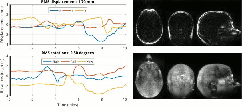
There is continual interest in pushing the boundaries of what can be achieved with MRI, especially regarding the spatial resolution of the images. At CUBRIC we are fortunate to have 4 state-of-the-art MR systems, including a very powerful 7T magnet (scanners in hospitals typically operate at 1.5T or 3T). This power enables us to acquire full 3D images of the brain with exceptionally high resolution (voxel sizes < 500 microns) - yet these high resolutions sitll require long scan times. It is easy to understand that during long scan times (up to 30 mins or more) you will probably move your head by a milimetre or two, even if you try to remain as still as possible - and with these very high resolution images this will still affect the achievable image quality.
Over the past few years - mainly while working at the EPFL in Switzerland - I have been developing a method to use rapid acquisitions of just the fat within the head (3D FatNavs) to allow tracking of very tiny head movements - which can then be corrected by post-processing of the raw data.
We are keen for other sites to start trying out 3D FatNavs - and we already have collaborators testing the seqeunces at different sites, both on Siemens and Philips platforms. If you are interested in collaborating, please contact me by email.
The set of Matlab tools which were developed to perform the whole retrospective correction pipeline can also be freely downloaded from the RetroMoCoBox Github page.
Imaging the brain at ultra-high resolution
In May 2016, we also demonstrated using 3D FatNavs to allow ultra-high resolution imaging at 7T, down to around 350 micron isotropic resolution of the whole brain. The full paper is Open Access and available from PLOS ONE - and you can also download the full datasets in NIFTI format from the Open Science Framework.
Manually segmented hippocampus 3D software rendering of a manually segmented hippocampus from an ultra-high resolution MRI scan. Read more in Federau and Gallichan, PLOS ONE 2016. |
The principle behind 3D FatNavs
A typical MRI image of the head at low resolution (2mm) might take around 30s to acquire:
But if we perform the same scan but at the frequency specific to fat rather than water, we obtain this image:
The fat within the head is primarily localized to the scalp, which results in an image which is sparse (i.e. most of the image is zero or close to zero). Sparsity is an important concept in signal processing, as sparse signals can be more easily compressed without losing information. The corresponding concept In MRI is that if we can represent our image in a sparse way, then we should also be able to acquire the data for our image much faster (via parallel imaging) while still maintaining a reasonable image quality.
Here is the same image, but acquired in just over 1 second (an effective acceleration factor of 28 is achieved by combining 4x4 GRAPPA acceleration with 6/8 partial Fourier in both phase-encoding directions):
The concept of the 3D FatNav is therefore to regularly acquire volumes such as that shown above (for example, a 'fat navigator' image could be acquired every 6 seconds during an MP2RAGE structural scan) and to track the small movements of the head which occurred during the whole scan by aligning these fat images. The motion information can then be used to retrospectively correct the raw data from the primary structural scan via post-processing.
Animated versions of figures from 3D FatNavs paper
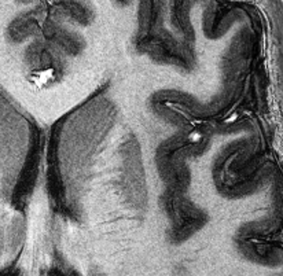
Figure 4 from Gallichan et al
A zoomed section of the full MP2RAGE volume acquired at 0.33x0.33x1.00 mm, showing the improvements following retrospective motion-correction using the 3D FatNavs
Figure 5 from Gallichan et al
A zoomed section of the GRETI2 volume from the same 0.33x0.33x1.00mm MP2RAGE dataset, with a minimum intensity projection taken over a 10 mm slab in the z-direction, showing the improvements following retrospective motion-correction using the 3D FatNavs
Figure 6 from Gallichan et al
Figure 6 from Gallichan et al
A zoomed section of the GRETI2 volume from the same 0.33x0.33x1.00mm MP2RAGE dataset, with a maximum intensity projection taken over the full 80 mm slab in the z-direction, showing the improvements following retrospective motion-correction using the 3D FatNavs
Figure 8 from Gallichan et al
Figure 8 from Gallichan et al
A zoomed section of the 0.6x0.6x0.6mm TSE volume showing the improvement following retrospective motion-correction using the 3D FatNavs
Teaching
Currently I teach introductory programming in Python to our MechEng and MedEng students, and I also teach MRI and more general medical imaging in the third year for the MedEng students.
Biography
Education and qualifications
- 2007: DPhil in Medical Physics. University of Oxford. Measuring Cerebral Blood Flow using ASL in MRI.
- 2003: Life Sciences Interface Doctoral Training Year. University of Oxford.
- 2002: MSci Physics with a European Language (German), University of Nottingham (exchange year at LMU Munich)
Career overview
- 2016 - present: Lecturer in Engineering with research at CUBRIC, Cardiff University
- 2011 - 2016: Senior Research Scientist, EPFL Lausanne, Switzerland
- 2009 - 2011: Post-doctoral Research Scientist, University Medical Center Freiburg, Germany
- 2007 - 2008: Post-doctoral Research Scientist, FMRIB Centre, University of Oxford
Supervisions
I am interested in supervising PhD students in the areas of:
- Motion-correction for MRI
- Novel MRI pulse sequence development
- Advanced MRI reconstruction methods
- Any interesting project in 'MR Physics'!
Current supervision

Contact Details
+44 29208 70045
Cardiff University Brain Research Imaging Centre, Maindy Road, Cardiff, CF24 4HQ
Queen's Buildings - East Building, Room E/3.24, 5 The Parade, Newport Road, Cardiff, CF24 3AA
Research themes
Specialisms
- Biomedical imaging
- Image processing
- Computational imaging
- Medical physics
- Medical devices