
Professor Derek Jones
(he/him)
FISMRM MBE FRSB DipIPSM
- Available for postgraduate supervision
Teams and roles for Derek Jones
Professor, Director of CUBRIC
Overview
CUBRIC's core mission is to advance brain imaging to improve cognitive, physical, and mental health across the lifespan in health and disease. Our vision is to harness the power of neuroimaging to better global health and quality of life. As the Director, I provide strategic guidance to the centre's pursuits.
My own research centres on optimizing non-invasive magnetic resonance imaging to extract quantitative insights into brain structure, in health (including in development and ageing), and in disease (including a range of neurological, psychiatric and oncological conditions).
You can find my publications, citations, h-index etc. at this Google Scholar link
You may find the journalistic narrative piece linked here useful in understanding what I do.
My recent work, predominantly in diffusion MRI, has focused on two extremes:
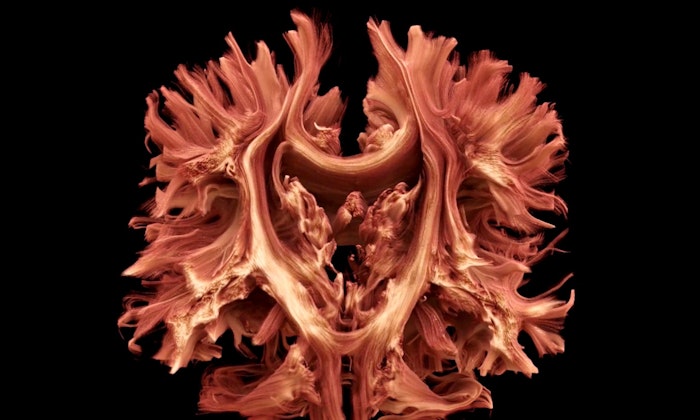
1. Exploiting ultra-strong imaging gradients
We're pushing the limits of tissue microstructural characterisation in white and grey matter using the Siemens Connectom scanner. The picture below shows my own brain (a still from the BBC news item that you can watch here).
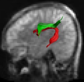
2. Developing diffusion MRI at low field
Working with the Bill and Melinda Gates Foundation, our group is developing the ability to reconstruct white matter pathways on low cost, low field, ultra-portable MRI machines. We are primarily targeting the ability to 'democratise MRI' allowing everyone, everyhere in the world benefit from the advantages of MRI.
The picture below shows reconstruction of language pathways at 64 mT.
This latter work aligns with my passion for democratising access to MRI in its broadest sense.
More generally, I'm interested in any techniques that provide a deeper understanding of microstructure in tissue - both in health (e.g. in neurodevelopment) and in disease.
Publication
2026
- Casella, C. et al. 2026. Differences in white matter detected by ex vivo 9.4 T MRI are associated with axonal changes in the R6/1 model of Huntington's disease. Neurobiology of Disease 220 107318. (10.1016/j.nbd.2026.107318)
- Merritt, K. et al., 2026. Inflammatory markers (IL-6 and CRP) in childhood and their association with brain structure and psychotic experiences in adulthood. Brain, Behavior, and Immunity 133 106247. (10.1016/j.bbi.2025.106247)
- Navarro-González, R. et al., 2026. Increased brain-age gap in young adults with psychotic experiences. Biological Psychiatry: Global Open Science 6 (2) 100643. (10.1016/j.bpsgos.2025.100643)
- Karat, B. G. et al., 2026. Microstructural variation of hippocampal substructures across childhood and adolescence quantified with high-gradient diffusion MRI. Communications Biology (10.1038/s42003-026-09622-x)
- Coveney, S. et al., 2026. Robust constrained weighted least squares for in vivo human cardiac diffusion kurtosis imaging. Magnetic Resonance in Medicine 95 (1), pp.220-233. (10.1002/mrm.70037)
- París, G. et al., 2026. Thermal noise lowers the accuracy of rotationally invariant harmonics of diffusion MRI data and their robustness to experimental variations. Magnetic Resonance in Medicine 95 (1), pp.204-219. (10.1002/mrm.70035)
- Paul, S. et al., 2026. Motion compensated spin echo cardiac diffusion tensor imaging in multiple cardiac phases using an ultrahigh gradient strength scanner. Journal of Cardiovascular Magnetic Resonance 102699. (10.1016/j.jocmr.2026.102699)
- Afzali, M. et al., 2026. ID#: 2202749 Multiphase systolic and diastolic cardiac diffusion tensor imaging using higher order motion compensation at 300 mT/m gradient strength [Abstract]. Journal of Cardiovascular Magnetic Resonance 28 (Supp.1), pp.4-5. 102213. (10.1016/j.jocmr.2025.102213)
- Ringshaw, J. E. et al., 2026. Feasibility and validity of ultra- low-field MRI for measurement of regional infant brain volumes in structures associated with antenatal maternal anemia. Human Brain Mapping 47 (1) e70443. (10.1002/hbm.70443)
- Karat, B. G. et al., 2026. Revisiting the interpretation of Axon diameter mapping using higher-order signal representations. Imaging Neuroscience 4 IMAG.a.1080. (10.1162/imag.a.1080)
2025
- McNabb, C. B. et al. 2025. Controlled antenatal thyroid screening study III: effects of gestational thyroid status on brain microstructure. The Journal of Clinical Endocrinology & Metabolism 110 (12), pp.3322-3330. (10.1210/clinem/dgaf277)
- Afzali, M. et al. 2025. Cardiac diffusion kurtosis imaging in the human heart in vivo using 300mT/m gradients. Magnetic Resonance in Medicine 94 (5), pp.2100-2112. (10.1002/mrm.30626)
- Cai, Q. et al. 2025. Decoding brain structure-function dynamics in health and in psychosis via an autoencoder. Scientific Reports 15 40052. (10.1038/s41598-025-24232-z)
- Silva, A. et al. 2025. Penetrance of neurodevelopmental copy number variants is associated with variations in cortical morphology. Biological Psychiatry: Cognitive Neuroscience and Neuroimaging 10 (10), pp.1093-1106. (10.1016/j.bpsc.2025.05.010)
- Harrison, J. R. et al. 2025. White matter microstructure in mid- to late adulthood is influenced by pathway-stratified polygenic risk for Alzheimer’s disease. Frontiers in Neuroscience 19 1638503. (10.3389/fnins.2025.1638503)
- Mason, A. J. C. et al., 2025. The effect of developmental trauma on brain structures involved in threat and memory processing and its relation to psychotic experiences in adulthood. Schizophrenia Bulletin: The Journal of Psychoses and Related Disorders sbaf184. (10.1093/schbul/sbaf184)
- Lena, B. et al., 2025. Repeatability and reproducibility of rapid T1 mapping of brain tissues at 64 mT: a multicentre study. Imaging Neuroscience 3 IMAG.a.916. (10.1162/IMAG.a.916)
- McCloskey, H. et al. 2025. Quantified head-ball impacts in soccer: a preliminary, prospective study. Neurotrauma Reports 6 (1), pp.928-943. (10.1177/2689288x251380145)
- Gulani, V. et al., 2025. Expanding access to mri: the role of all-purpose mid-field and 1.5-t scanners. Radiology 316 (3) e251406. (10.1148/radiol.251406)
- Urbini, N. et al., 2025. The cognitive cerebellum: linking microstructure to cognitive functions in a healthy population. NeuroImage 317 121356. (10.1016/j.neuroimage.2025.121356)
- Bonham, K. S. et al., 2025. Codevelopment of gut microbial metabolism and visual neural circuitry over human infancy. mBio 16 (8) e00835-25. (10.1128/mbio.00835-25)
- Ringshaw, J. E. et al., 2025. Iron deficiency anaemia in mothers and infants with high inflammatory burden: Prevalence and profile in a South African birth cohort. PLOS Global Public Health 5 (7) e0004174. (10.1371/journal.pgph.0004174)
- Schiavi, S. et al., 2025. MRI and non-MRI quantifiable neuroanatomical and functional parameters are useful for tractography. Brain Structure and Function 230 83. (10.1007/s00429-025-02932-6)
- Borra, E. et al., 2025. Brain connectivity: complex, not chaotic. Brain Structure and Function 230 (5) 77. (10.1007/s00429-025-02943-3)
- Coveney, S. et al., 2025. Outlier detection in cardiac diffusion tensor imaging: Shot rejection or robust fitting?. Medical Image Analysis 101 103386. (10.1016/j.media.2024.103386)
- Cicimen, A. G. et al., 2025. Image quality transfer of diffusion MRI guided By high-resolution structural MRI. Presented at: CDMRI 2024 Marrakesh, Morocco 06 October 2024. Published in: Chamberland, M. et al., Computational Diffusion MRI. Lecture Notes in Computer Science Springer Nature Switzerland. , pp.106-118. (10.1007/978-3-031-86920-4_10)
- Genc, S. et al. 2025. MRI signatures of cortical microstructure in human development align with oligodendrocyte cell-type expression. Nature Communications 16 3317. (10.1038/s41467-025-58604-w)
- Molendowska, M. et al. 2025. Giving the prostate the boost it needs: Spiral diffusion MRI using a high-performance whole-body gradient system for high b-values at short echo times. Magnetic Resonance in Medicine 93 (3), pp.1256-1272. (10.1002/mrm.30351)
- Canales-Rodríguez, E. J. et al., 2025. A diffusion MRI model for random walks confined on cylindrical surfaces: towards non-invasive quantification of myelin sheath radius. Frontiers in Physics 13 1516630. (10.3389/fphy.2025.1516630)
- McNabb, C. B. et al. 2025. WAND: A multi-modal dataset integrating advanced MRI, MEG, and TMS for multi-scale brain analysis. Scientific Data 12 220. (10.1038/s41597-024-04154-7)
- Jones, D. K. et al. 2025. Low field, high impact: Democratizing MRI for clinical and research innovation. BJR Open 7 (1) tzaf022. (10.1093/bjro/tzaf022)
- Chamberland, M. et al., 2025. Methods and statistics for diffusion MRI tractometry. In: Dell'acqua, F. , Descoteaux, M. and Leemans, A. eds. Handbook of Diffusion MR Tractography: Imaging Methods, Biophysical Models, Algorithms and Applications. Elsevier. , pp.439-450. (10.1016/B978-0-12-818894-1.00023-9)
- Aird-Rossiter, C. et al. 2025. Decoding Gray Matter: large-scale analysis of brain cell morphometry to inform microstructural modeling of diffusion MR signals. Communications Biology 9 138. (10.1038/s42003-025-09353-5)
- Hiscox, L. V. et al. 2025. MR elastography reveals lower hippocampal stiffness in middle-aged APOE ε4 carriers without cognitive impairment. Presented at: International Society for Magnetic Resonance in Medicine (ISMRM) Hawaii, USA. 10-15 May 2025. Proceedings of the International Society for Magnetic Resonance in Medicine - Scientific Meeting and Exhibition. , pp.0791. (10.58530/2025/0791)
2024
- Wang, H. et al. 2024. Improving high-frequency details in cerebellum for brain MRI super-resolution. Presented at: Conference on ICT Solutions for eHealth (ICTS4eHealth 2024) Paris, France 26 - 29 June 2024. 2024 IEEE Symposium on Computers and Communications (ISCC). IEEE. , pp.1-7. (10.1109/ISCC61673.2024.10733580)
- Genc, S. et al. 2024. Developmental differences in canonical cortical networks: insights from microstructure-informed tractography. Network Neuroscience 8 (3), pp.946-964. (10.1162/netn_a_00378)
- Afzali, M. et al. 2024. In vivo diffusion MRI of the human heart using a 300 mT/m gradient system. Magnetic Resonance in Medicine 92 (3), pp.1022-1034. (10.1002/mrm.30118)
- Margolis, E. T. et al., 2024. Longitudinal effects of prenatal alcohol exposure on visual neurodevelopment over infancy. Developmental Psychology 60 (9), pp.1673–1698. (10.1037/dev0001727)
- MacIver, C. L. et al. 2024. White matter microstructural changes using ultra-strong diffusion gradient MRI in adult-onset idiopathic focal cervical dystonia. Neurology 103 (4) e209695. (10.1212/WNL.0000000000209695)
- Molendowska, M. et al. 2024. Diffusion MRI in prostate cancer with ultra-strong whole body gradients. NMR in Biomedicine (10.1002/nbm.5229)
- Ioakeimidis, V. et al. 2024. Protocol for a randomised controlled unblinded feasibility trial of HD-DRUM, a rhythmic movement training application for cognitive and motor symptoms in people with Huntington’s disease. BMJ Open 14 (7) e082161. (10.1136/bmjopen-2023-082161)
- Canales-Rodríguez, E. J. et al., 2024. Pore size estimation in axon-mimicking microfibers with diffusion-relaxation MRI. Magnetic Resonance in Medicine 91 (6), pp.2579-2596. (10.1002/mrm.29991)
- Planchuelo-Gómez, Á. et al. 2024. Optimisation of quantitative brain diffusion-relaxation MRI acquisition protocols with physics-informed machine learning. Medical Image Analysis 94 103134. (10.1016/j.media.2024.103134)
- Qiu, Z. et al., 2024. Self-calibrated subspace reconstruction for multidimensional MR fingerprinting for simultaneous relaxation and diffusion quantification. Magnetic Resonance in Medicine 91 (5), pp.1978-1993. (10.1002/mrm.29969)
- Scholz, A. et al. 2024. Controlled antenatal thyroid screening study III: Effects of gestational thyroid status on adolescent brain morphology. The Journal of Clinical Endocrinology & Metabolism (10.1210/clinem/dgae338)
- Engel, M. et al. 2024. Maximising SNR per unit time in diffusion MRI with multiband T-Hex spirals. Magnetic Resonance in Medicine 91 (4), pp.1323-1336. (10.1002/mrm.29953)
- Jones, D. K. 2024. Commentary for “Environmental Sustainability and MRI: Challenges, Opportunities, and a Call for Action”. Journal of Magnetic Resonance Imaging 59 (4), pp.1168-1169. (10.1002/jmri.29118)
- Uhl, Q. et al., 2024. Quantifying human gray matter microstructure using neurite exchange imaging (NEXI) and 300 mT/m gradients. Imaging Neuroscience 2 , pp.1-19. (10.1162/imag_a_00104)
2023
- Endt, S. et al. 2023. In vivo myelin water quantification using diffusion–relaxation correlation MRI: A comparison of 1D and 2D methods. Applied Magnetic Resonance 54 , pp.1571-1588. (10.1007/s00723-023-01584-1)
- Davies Jenkins, C. W. et al., 2023. Practical considerations of diffusion-weighted MRS with ultra-strong diffusion gradients. Frontiers in Neuroscience 17 1258408. (10.3389/fnins.2023.1258408)
- MacIver, C. et al. 2023. Macro- and micro-structural Insights into primary dystonia A UK Biobank study. Journal of Neurology (10.1007/s00415-023-12086-2)
- Ioakeimidis, V. et al. 2023. Protocol for a randomised controlled feasibility trial of HD-DRUM, a rhythmic movement training application for cognitive and motor symptoms in people with Huntington's disease. [Online].medRxiv: medRxiv. (10.1101/2023.11.15.23298581)Available at: https://doi.org/10.1101/2023.11.15.23298581.
- Raven, E. et al. 2023. In vivo evidence of microstructural hypo-connectivity of brain white matter in 22q11.2 deletion syndrome. Molecular Psychiatry 28 , pp.4342-4352. (10.1038/s41380-023-02178-w)
- Merritt, K. et al., 2023. The impact of cumulative obstetric complications and childhood trauma on brain volume in young people with psychotic experiences. Molecular Psychiatry 28 , pp.3688-3697. (10.1038/s41380-023-02295-6)
- Kleban, E. , Jones, D. and Tax, C. 2023. The impact of head orientation with respect to B0 on diffusion tensor MRI measures. Imaging Neuroscience 1 , pp.1-17. (10.1162/imag_a_00012)
- Harrison, J. R. et al. 2023. Pathway-specific polygenic scores for Alzheimer's disease are associated with changes in brain structure in younger and older adults. Brain Communications 5 (5) fcad229. (10.1093/braincomms/fcad229)
- Barakovic, M. et al. 2023. Estimating axon radius using diffusion-relaxation MRI: calibrating a surface-based relaxation model with histology. Frontiers in Neuroscience 17 1209521. (10.3389/fnins.2023.1209521)
- Wang, H. et al. 2023. A skewed loss function for correcting predictive bias in brain age prediction. IEEE Transactions on Medical Imaging 42 (6), pp.1577-1589. (10.1109/TMI.2022.3231730)
- Genc, S. et al. 2023. Novel insights into axon diameter and myelin content in late childhood and adolescence. Cerebral Cortex 33 (10), pp.6435-6448. (10.1093/cercor/bhac515)
- Tax, C. M. W. et al. 2023. Ultra-strong diffusion-weighted MRI reveals cerebellar grey matter abnormalities in movement disorders. NeuroImage: Clinical 38 103419. (10.1016/j.nicl.2023.103419)
- Ward, I. L. et al. 2023. White matter microstructure in face and body networks predicts facial expression and body posture perception across development. Human Brain Mapping 44 (6), pp.2307-2322. (10.1002/hbm.26211)
- Warner, W. et al., 2023. Temporal Diffusion Ratio (TDR) for imaging restricted diffusion: optimisation and pre-clinical demonstration. NeuroImage 269 119930. (10.1016/j.neuroimage.2023.119930)
- Dimitriadis, S. I. et al. 2023. Genetic risk for schizophrenia is associated with increased proportion of indirect connections in brain networks revealed by a semi-metric analysis: evidence from population sample stratified for polygenic risk. Cerebral Cortex 33 (6), pp.2997-3011. (10.1093/cercor/bhac256)
- Messaritaki, E. et al. 2023. Increased structural connectivity in high schizotypy. Network Neuroscience 7 (1), pp.213-233. (10.1162/netn_a_00279)
2022
- Afzali, M. et al. 2022. MR Fingerprinting with b-tensor encoding for simultaneous quantification of relaxation and diffusion in a single scan. Magnetic Resonance in Medicine 88 (5), pp.2043-2057. (10.1002/mrm.29352)
- MacIver, C. L. et al. 2022. Structural magnetic resonance imaging in dystonia: A systematic review of methodological approaches and findings. European Journal of Neurology 29 (11), pp.3418-3448. (10.1111/ene.15483)
- Mirza-Davies, A. et al. 2022. The impact of genetic risk for Alzheimer’s disease on the structural brain networks of young adults. Frontiers in Neuroscience 16 987677. (10.3389/fnins.2022.987677)
- Howard, A. F. D. et al., 2022. Estimating axial diffusivity in the NODDI model. NeuroImage 262 119535. (10.1016/j.neuroimage.2022.119535)
- Shastin, D. et al. 2022. Surface-based tracking for short association fibre tractography. NeuroImage 260 119423. (10.1016/j.neuroimage.2022.119423)
- Metzler-Baddeley, C. et al. 2022. HD-DRUM – a novel computerised drumming training for movement and cognitive abilities in people with Huntington’s disease – app development and protocol of a randomised controlled feasibility study. Presented at: EHDN 2022 Plenary Meeting Bologna, Italy 16-18 September 2022. Vol. 93.Vol. S1. , pp.A100-A101. (10.1136/jnnp-2022-ehdn.267)
- Afzali, M. et al. 2022. Cumulant expansion with localization: a new representation of the diffusion MRI signal. Frontiers in Neuroimaging 1 958680. (10.3389/fnimg.2022.958680)
- Casella, C. et al. 2022. Mutation-related magnetization-transfer, not axon density, drives white matter differences in premanifest Huntington's disease: Evidence from in vivo ultra-strong gradient MRI. Human Brain Mapping 43 (11), pp.3439-3460. (10.1002/hbm.25859)
- Fan, Q. et al., 2022. Mapping the human connectome using diffusion MRI at 300 mT/m gradient strength: methodological advances and scientific impact. NeuroImage 254 118958. (10.1016/j.neuroimage.2022.118958)
- Aja-Fernández, S. et al., 2022. Anisotropy measure from three diffusion-encoding gradient directions. Magnetic Resonance Imaging 88 , pp.38-43. (10.1016/j.mri.2022.01.014)
- Molendowska, M. et al. 2022. Physiological effects of human body imaging with 300 mT/m gradients. Magnetic Resonance in Medicine 87 (5), pp.2512-2520. (10.1002/mrm.29118)
- Garcia-Hernandez, R. et al., 2022. Mapping microglia and astrocyte activation in vivo using diffusion MRI. Science Advances 8 (21) eabq2923. (10.1126/sciadv.abq2923)
- Baker, R. R. et al., 2022. Image-guided magnetic thermoseed navigation and tumor ablation using a magnetic resonance imaging system. Advanced Science 9 (12) 2105333. (10.1002/advs.202105333)
- Schiavi, S. et al., 2022. Bundle myelin fraction (BMF) mapping of different white matter connections using microstructure informed tractography. NeuroImage 249 118922. (10.1016/j.neuroimage.2022.118922)
- Afzali, M. et al., 2022. Quantification of tissue microstructure using tensor-valued diffusion encoding: brain and body. Frontiers of Physics 10 809133. (10.3389/fphy.2022.809133)
2021
- Dimitriadis, S. I. et al. 2021. Global brain flexibility during working memory is reduced in a high genetic risk group for schizophrenia. Biological Psychiatry: Cognitive Neuroscience and Neuroimaging 6 (12), pp.1176-1184. (10.1016/j.bpsc.2021.01.007)
- Casella, C. et al. 2021. Mutation-related apparent myelin, not axon density, drives white matter differences in premanifest Huntington's disease: evidence from in vivo ultra-strong gradient MRI. bioRxiv (10.1101/2021.11.29.469517)
- Dimitriadis, S. I. et al. 2021. Genetic risk for schizophrenia is associated with altered visually-induced gamma band activity: evidence from a population sample stratified polygenic risk. Translational Psychiatry 11 592. (10.1038/s41398-021-01678-z)
- De Luca, A. et al., 2021. On the generalizability of diffusion MRI signal representations across acquisition parameters, sequences and tissue types: chronicles of the MEMENTO challenge. NeuroImage 240 118367. (10.1016/j.neuroimage.2021.118367)
- Chamberland, M. et al. 2021. Detecting microstructural deviations in individuals with deep diffusion MRI tractometry. Nature Computational Science 1 , pp.598-606. (10.1038/s43588-021-00126-8)
- Chamberland, M. et al. 2021. Beyond lesion-load: tractometry-based metrics for characterizing white matter lesions within fibre pathways. Presented at: MICCAI 2020 International Workshop on Computational Diffusion MRI (CDMRI 2020) Virtual 8 October 2020. Published in: Gyori, N. et al., Computational Diffusion MRI: International MICCAI Workshop, Lima, Peru, October 2020. Mathematics and Visualization , pp.227-237. (10.1007/978-3-030-73018-5_18)
- Dimitriadis, S. I. , Messaritaki, E. and Jones, D. K. 2021. The impact of graph construction scheme and community detection algorithm on the reliability of community and hub identification in structural brain networks. Human Brain Mapping 42 (13), pp.4261-4280. (10.1002/hbm.25545)
- Pieciak, T. et al., 2021. Q-Space quantitative diffusion MRI measures using a stretched-exponential representation. Presented at: International MICCAI Workshop Lima, Peru Oct 2020. Published in: Gyori, N. et al., Computational Diffusion MRI. Mathematics and Visualization Springer. , pp.121-133. (10.1007/978-3-030-73018-5_10)
- Zappala, S. et al. 2021. Full-field MRI measurements of in-vivo positional brain shift reveal the significance of intra-cranial geometry and head orientation for stereotactic surgery. Scientific Reports 11 (1) 17684. (10.1038/s41598-021-97150-5)
- Afzali, M. et al. 2021. SPHERIOUSLY? The challenges of estimating sphere radius non-invasively in the human brain from diffusion MRI. NeuroImage 237 118183. (10.1016/j.neuroimage.2021.118183)
- Tax, C. M. W. et al. 2021. Measuring compartmental T2-orientational dependence in human brain white matter using a tiltable RF coil and diffusion-T2 correlation MRI. NeuroImage 236 117967. (10.1016/j.neuroimage.2021.117967)
- Winter, M. et al. 2021. Tract-specific MRI measures explain learning and recall differences in multiple sclerosis. Brain Communications 3 (2) fcab065. (10.1093/braincomms/fcab065)
- Afzali, M. et al., 2021. Computing the orientational-average of diffusion-weighted MRI signals: a comparison of different techniques. Scientific Reports 11 14345. (10.1038/s41598-021-93558-1)
- Messaritaki, E. et al. 2021. Predicting MEG resting-state functional connectivity using microstructural information. Network Neuroscience 5 (2), pp.477-504. (10.1162/netn_a_00187)
- Henriques, R. N. et al., 2021. Towards more robust and reproducible diffusion kurtosis imaging. Magnetic Resonance in Medicine 86 (3), pp.1600-1613. (10.1002/mrm.28730)
- Huang, C. et al., 2021. Validating pore size estimates in a complex microfibre environment on a human MRI system. Magnetic Resonance in Medicine 86 (3), pp.1514-1530. (10.1002/mrm.28810)
- Lipp, I. et al. 2021. Predictors of training-related improvement in visuomotor performance in patients with multiple sclerosis: a behavioural and MRI study. Multiple Sclerosis 7 (7), pp.1088-1101. (10.1177/1352458520943788)
- Aja-Fernández, S. , Tristán-Vega, A. and Jones, D. K. 2021. Apparent propagator anisotropy from single-shell diffusion MRI acquisitions. Magnetic Resonance in Medicine 85 (5), pp.2869-2881. (10.1002/mrm.28620)
- Veraart, J. et al., 2021. The variability of MR axon radii estimates in the human white matter. Human Brain Mapping 42 (7), pp.2201-2213. (10.1002/hbm.25359)
- Yeh, C. et al., 2021. Mapping structural connectivity using diffusion MRI : challenges and opportunities. Journal of Magnetic Resonance Imaging 53 (6), pp.1666-1682. (10.1002/jmri.27188)
- Casella, C. et al. 2021. Multi-compartment analysis of the complex gradient-echo signal quantifies myelin breakdown in premanifest Huntington's disease. NeuroImage: Clinical 30 102658. (10.1016/j.nicl.2021.102658)
- Barakovic, M. et al. 2021. Resolving bundle-specific intra-axonal T2 values within a voxel using diffusion-relaxation tract-based estimation. NeuroImage 227 117617. (10.1016/j.neuroimage.2020.117617)
- Tax, C. M. W. et al. 2021. Magnetic resonance imaging of T2 - and diffusion snisotropy using a tiltable receive coil. Presented at: Visualization and Processing of Anisotropy in Imaging, Geometry, and Astronomy Dagstuhl, Germany 28 Oct–2 Nov 2018. Published in: Ozarslan, E. et al., Anisotropy Across Fields and Scales. Mathematics and Visualization Springer. , pp.247-262. (10.1007/978-3-030-56215-1_12)
- de Almeida Martins, J. P. et al., 2021. Computing and visualising intra-voxel orientation-specific relaxation-diffusion features in the human brain. Human Brain Mapping 42 (2), pp.310-328. (10.1002/hbm.25224)
- Guo, F. et al., 2021. The effect of gradient nonlinearities on fiber orientation estimates from spherical deconvolution of diffusion MRI data. Human Brain Mapping 42 (2), pp.367-383. (10.1002/hbm.25228)
- Koller, K. et al. 2021. MICRA: Microstructural Image Compilation with Repeated Acquisitions. NeuroImage 225 117406. (10.1016/j.neuroimage.2020.117406)
- Afzali, M. et al., 2021. The sensitivity of diffusion MRI to microstructural properties and experimental factors. Journal of Neuroscience Methods 347 108951. (10.1016/j.jneumeth.2020.108951)
- Gholam, J. A. et al. 2021. aDWI-BIDS: advanced diffusion weighted imaging metadata for the brain imaging data structure. Presented at: ISMRM & SMRT Annual Meeting & Exhibition Virtual 15-20 May 2021.
2020
- Ning, L. et al., 2020. Cross-scanner and cross-protocol multi-shell diffusion MRI data harmonization: algorithms and result. NeuroImage 221 117128. (10.1016/j.neuroimage.2020.117128)
- Pizzolato, M. et al., 2020. Acquiring and predicting multidimensional diffusion (MUDI) data: an open challenge. Presented at: MICCAI Workshop Shenzhen, China Oct 2019. Published in: Bonet-Carne, E. et al., Computational Diffusion MRI. Mathematics and Visualization Springer. , pp.195-208. (10.1007/978-3-030-52893-5_17)
- Rafael-Patino, J. et al., 2020. DWI simulation-assisted machine learning models for microstructure estimation. Presented at: MICCAI Workshop Shenzhen, China Oct 2019. Published in: Bonet-Carne, E. et al., Computational Diffusion MRI. Mathematics and Visualization Springer. , pp.125-134. (10.1007/978-3-030-52893-5_11)
- Casella, C. et al. 2020. Drumming motor sequence training induces apparent myelin remodelling in Huntington’s disease: a longitudinal diffusion MRI and quantitative magnetization transfer study. Journal of Huntington's Disease 9 (3), pp.303-320. (10.3233/JHD-200424)
- Afzali, M. , Aja-Fernandez, S. and Jones, D. K. 2020. Direction-averaged diffusion-weighted MRI signal using different axisymmetric B-tensor encoding schemes. Magnetic Resonance in Medicine 84 (3), pp.1579-1591. (10.1002/mrm.28191)
- Casella, C. et al. 2020. A critical review of white matter changes in Huntington’s disease. Movement Disorders 35 (8), pp.1302-1311. (10.1002/mds.28109)
- Sharp, T. H. et al., 2020. Population neuroimaging: generation of a comprehensive data resource within the ALSPAC pregnancy and birth cohort. Wellcome Open Research 5 203. (10.12688/wellcomeopenres.16060.1)
- Kleban, E. et al. 2020. Strong diffusion gradients allow the separation of intra- and extra-axonal gradient-echo signals in the human brain. NeuroImage 217 116793. (10.1016/j.neuroimage.2020.116793)
- Genc, S. et al. 2020. Impact of b-value on estimates of apparent fibre density. Human Brain Mapping 41 (10), pp.2583-2595. (10.1002/hbm.24964)
- Tax, C. et al. 2020. The dot-compartment revealed? Diffusion MRI with ultra-strong gradients and spherical tensor encoding in the living human brain. NeuroImage 210 116534. (10.1016/j.neuroimage.2020.116534)
- Lipp, I. et al. 2020. Tractography in the presence of multiple sclerosis lesions. NeuroImage 209 116471. (10.1016/j.neuroimage.2019.116471)
- Postans, M. et al. 2020. Uncovering a role for the dorsal hippocampal commissure in recognition memory. Cerebral Cortex 30 (3), pp.1001-1015. (10.1093/cercor/bhz143)
- Poudel, G. R. et al., 2020. Network diffusion modeling predicts neurodegeneration in traumatic brain injury. Annals of Clinical and Translational Neurology 7 (3), pp.270-279. (10.1002/acn3.50984)
- Martins, J. P. d. A. et al., 2020. Transferring principles of solid-state and Laplace NMR to the field of in vivo brain MRI. Magnetic Resonance 1 , pp.27-43. (10.5194/mr-1-27-2020)
- Rudrapatna, U. et al., 2020. A comparative study of gradient nonlinearity correction strategies for processing diffusion data obtained with ultra-strong-gradient MRI scanner. Magnetic Resonance in Medicine 85 (2), pp.1104-1113. (10.1002/mrm.28464)
- Veraart, J. et al., 2020. Noninvasive quantification of axon radii using diffusion MRI. eLife 9 e49855. (10.7554/eLife.49855)
- Chamberland, M. et al. 2020. Tractometry-based anomaly detection for single-subject white matter analysis. Presented at: Medical Imaging with Deep Learning (MIDL 2020) Montréal, Canada 6-9 July 2020.
- Harrison, J. R. et al. 2020. Imaging Alzheimer's genetic risk using Diffusion MRI: a systematic review. NeuroImage: Clinical 27 102359. (10.1016/j.nicl.2020.102359)
2019
- Drakesmith, M. et al. 2019. Estimating axon conduction velocity in vivo from microstructural MRI. NeuroImage 203 116186. (10.1016/j.neuroimage.2019.116186)
- Chamberland, M. et al. 2019. Dimensionality reduction of diffusion MRI measures for improved tractometry of the human brain. NeuroImage 200 , pp.89-100. (10.1016/j.neuroimage.2019.06.020)
- Messaritaki, E. , Dimitriadis, S. I. and Jones, D. K. 2019. Optimization of graph construction can significantly increase the power of structural brain network studies. NeuroImage 199 , pp.495-511. (10.1016/j.neuroimage.2019.05.052)
- Lipp, I. et al. 2019. Comparing MRI metrics to quantify white matter microstructural damage in multiple sclerosis. Human Brain Mapping 40 (10), pp.2917-2932. (10.1002/hbm.24568)
- Tax, C. M. W. et al. 2019. Cross-scanner and cross-protocol diffusion MRI data harmonisation: A benchmark database and evaluation of algorithms. NeuroImage 195 , pp.285-299. (10.1016/j.neuroimage.2019.01.077)
- Afzali Deligani, M. et al. 2019. Comparison of different tensor encoding combinations in microstructural parameter estimation. Presented at: IEEE International Symposium on Biomedical Imaging Venice, Italy 8-11 Apr 2019. 2019 IEEE 16th International Symposium on Biomedical Imaging (ISBI 2019). IEEE. , pp.1471-1474. (10.1109/ISBI.2019.8759100)
- Chamberland, M. et al. 2019. Obtaining representative core streamlines for white matter tractometry of the human brain. Presented at: International MICCAI Workshop Granada, Spain Sep 2018. Published in: Bonet-Carne, E. et al., Computational Diffusion MRI. Mathematics and Visualization Cham: Springer. , pp.359-366. (10.1007/978-3-030-05831-9_28)
- Ning, L. et al., 2019. Muti-shell diffusion MRI harmonisation and enhancement challenge (MUSHAC): Progress and results. Presented at: MICCAI 2018 Granada, Spain 16-20 September 2018. Published in: Bonet-Carne, E. et al., Computational Diffusion MRI. Vol. 1.Mathematics and Visualization Cham: Springer. , pp.217-224. (10.1007/978-3-030-05831-9_18)
- Bourbon Teles, J. et al., 2019. Myelin breakdown in human Huntington's disease: multi-modal evidence from diffusion MRI and quantitative magnetization transfer. Neuroscience 403 , pp.79-92. (10.1016/j.neuroscience.2017.05.042)
- Imms, P. et al., 2019. The structural connectome in traumatic brain injury: A meta-analysis of graph metrics. Neuroscience and Biobehavioral Reviews 99 , pp.128-137. (10.1016/j.neubiorev.2019.01.002)
- Metzler-Baddeley, C. et al. 2019. Sex-specific effects of central adiposity and inflammatory markers on limbic microstructure. NeuroImage 189 , pp.793-803. (10.1016/j.neuroimage.2019.02.007)
- Lancaster, T. M. et al. 2019. Structural and functional neuroimaging of polygenic risk for schizophrenia: a recall-by-genotype-based approach. Schizophrenia Bulletin 45 (2), pp.405-414. (10.1093/schbul/sby037)
- David, S. et al., 2019. The Superoanterior Fasciculus (SAF): a novel white matter pathway in the human brain?. Frontiers in Neuroanatomy 13 , pp.-. 24. (10.3389/fnana.2019.00024)
- Drakesmith, M. et al. 2019. Genetic risk for schizophrenia and developmental delay is associated with shape and microstructure of midline white-matter structures. Translational Psychiatry 9 (1) 102. (10.1038/s41398-019-0440-7)
- Metzler-Baddeley, C. et al. 2019. Fornix white matter glia damage causes hippocampal gray matter damage during age-dependent limbic decline. Scientific Reports 9 1060. (10.1038/s41598-018-37658-5)
- Fonville, L. et al., 2019. MRI indices of cortical development in young people with psychotic experiences: influence of genetic risk and persistence of symptoms. Schizophrenia Bulletin 45 (1), pp.169-179. sbx195. (10.1093/schbul/sbx195)
- Hyde, C. et al., 2019. White matter organization in developmental coordination disorder: a pilot study exploring the added value of constrained spherical deconvolution. NeuroImage: Clinical 21 101625. (10.1016/j.nicl.2018.101625)
2018
- Chamberland, M. , Tax, C. M. W. and Jones, D. K. 2018. Meyer's loop tractography for image-guided surgery depends on imaging protocol and hardware. NeuroImage: Clinical 20 , pp.458-465. (10.1016/j.nicl.2018.08.021)
- Messaritaki, E. et al. 2018. Improving the predictions of computational models of convection-enhanced drug delivery by accounting for diffusion non-Gaussianity. Frontiers in Neurology 9 1092. (10.3389/fneur.2018.01092)
- Jones, D. K. et al. 2018. Microstructural imaging of the human brain with a 'super-scanner': 10 key advantages of ultra-strong gradients for diffusion MRI. NeuroImage 182 , pp.8-38. (10.1016/j.neuroimage.2018.05.047)
- Molina-Romero, M. et al., 2018. A diffusion model-free framework with echo time dependence for free-water elimination and brain tissue microstructure characterization. Magnetic Resonance in Medicine 80 (5), pp.2155-2172. (10.1002/mrm.27181)
- Caeyenberghs, K. et al. 2018. Evidence for training-dependent structural neuroplasticity in brain-injured patients: A critical review. Neurorehabilitation and Neural Repair 32 (2), pp.99-114. (10.1177/1545968317753076)
2017
- Chamberland, M. et al. 2017. Interactive computation and visualization of structural connectomes in real-time. Presented at: CNI 2017: International Workshop on Connectomics in Neuroimaging Quebec City, QC, Canada 14 September 2017. Published in: Wu, G. et al., Connectomics in NeuroImaging. Vol. 10511.Lecture Notes in Computer Science Cham, Switzerland: Springer Verlag. , pp.35-41. (10.1007/978-3-319-67159-8_5)
- Metzler-Baddeley, C. et al. 2017. Dynamics of white matter plasticity underlying working memory training: Multi-modal evidence from diffusion MRI and relaxometry. Journal of Cognitive Neuroscience 29 (9), pp.1509-1520. (10.1162/jocn_a_01127)
- Dimitriadis, S. et al., 2017. Improving the reliability of network metrics in structural brain networks by integrating different network weighting strategies into a single graph. Frontiers in Neuroscience 11 694. (10.3389/fnins.2017.00694)
2016
- Christiansen, K. et al. 2016. Topographic separation of fornical fibers associated with the anterior and posterior hippocampus in the human brain: an MRI-diffusion study. Brain and Behavior 7 (1)(10.1002/brb3.604)
- Hunt, B. A. E. et al., 2016. Relationships between cortical myeloarchitecture and electrophysiological networks. Proceedings of the National Academy of Sciences 113 (47), pp.13510-13515. (10.1073/pnas.1608587113)
- Rok, B. et al., 2016. Global efficiency of structural networks mediates cognitive control in Mild Cognitive Impairment. Frontiers in Aging Neuroscience 8 292. (10.3389/fnagi.2016.00292)
- De Santis, S. et al. 2016. T1 relaxometry of crossing fibres in the human brain. NeuroImage 141 , pp.133-142. (10.1016/j.neuroimage.2016.07.037)
- Gómez, P. A. et al., 2016. Simultaneous parameter mapping, modality synthesis, and anatomical labeling of the brain with MR fingerprinting. Presented at: MICCAI 2016: nternational Conference on Medical Image Computing and Computer-Assisted Intervention Athens, Greece 17-21 October 2016. Published in: Ourselin, S. et al., Medical Image Computing and Computer-Assisted Intervention -- MICCAI 2016: 19th International Conference, Athens, Greece, October 17-21, 2016, Proceedings, Part III. Vol. 9902.Lecture Notes in Computer Science Cham: Springer. , pp.579-586. (10.1007/978-3-319-46726-9_67)
- Drakesmith, M. et al. 2016. Volumetric, relaxometric and diffusometric correlates of psychotic experiences in a non-clinical sample of young adults. NeuroImage: Clinical 12 , pp.550-558. (10.1016/j.nicl.2016.09.002)
- Steventon, J. et al. 2016. Longitudinal in vivo MRI in a Huntington's disease mouse model: global atrophy in the absence of white matter microstructural damage. Scientific Reports 6 32423. (10.1038/srep32423)
- Keedwell, P. A. et al., 2016. Subgenual cingulum microstructure supports control of emotional conflict. Cerebral Cortex 26 (6), pp.2850-2862. (10.1093/cercor/bhw030)
- Metzler-Baddeley, C. et al. 2016. Longitudinal data on cortical thickness before and after working memory training. Data in Brief 7 , pp.1143-1147. (10.1016/j.dib.2016.03.090)
- Steventon, J. et al. 2016. Robust MR-based approaches to quantifying white matter structure and structure/function alterations in Huntington's disease. Journal of Neuroscience Methods 265 , pp.2-12. (10.1016/j.jneumeth.2015.08.027)
- Drakesmith, M. et al. 2016. Mediation of psychosis risk factors by white-matter microstructure in young adults with psychotic experiences. JAMA Psychiatry 73 (4), pp.396-406. (10.1001/jamapsychiatry.2015.3375)
- Metzler-Baddeley, C. et al. 2016. Task complexity and location specific changes of cortical thickness in executive and salience networks after working memory training. NeuroImage 130 , pp.48-62. (10.1016/j.neuroimage.2016.01.007)
- Caeyenberghs, K. et al., 2016. Dynamics of the human structural connectome underlying working memory training. Journal of Neuroscience 36 (14), pp.4056-4066. (10.1523/JNEUROSCI.1973-15.2016)
- Dimitriadis, S. et al. 2016. A prolonged maturational time course in brain development for cortical processing of temporal modulations. Clinical Neurophysiology 127 (2), pp.994-998. (10.1016/j.clinph.2015.09.001)
- De Santis, S. , Jones, D. K. and Roebroeck, A. 2016. Including diffusion time dependence in the extra-axonal space improves in vivo estimates of axonal diameter and density in human white matter. NeuroImage 130 , pp.91-103. (10.1016/j.neuroimage.2016.01.047)
- Bracht, T. K. et al. 2016. Myelination of the right parahippocampal cingulum is associated with physical activity in young healthy adults. Brain Structure and Function 221 (9), pp.4537-48. (10.1007/s00429-016-1183-3)
- Rees, D. A. et al. 2016. White matter microstructure and cognitive function in young women with polycystic ovary syndrome. Journal of Clinical Endocrinology & Metabolism 101 (1), pp.314-323. (10.1210/jc.2015-2318)
- Ecker, C. et al., 2016. Relationship Between Cortical Gyrification, White Matter Connectivity, and Autism Spectrum Disorder. Cerebral Cortex 26 (7), pp.3297-3309. (10.1093/cercor/bhw098)
2015
- De Santis, S. et al. 2015. Resolving relaxometry and diffusion properties within the same voxel in the presence of crossing fibres by combining inversion recovery and diffusion-weighted acquisitions. Magnetic Resonance in Medicine 75 (1), pp.372-380. (10.1002/mrm.25644)
- Drakesmith, M. et al. 2015. Overcoming the effects of false positives and threshold bias in graph theoretical analyses of neuroimaging data. NeuroImage 118 , pp.313-333. (10.1016/j.neuroimage.2015.05.011)
- Hodgetts, C. J. et al. 2015. Dissociable roles of the inferior longitudinal fasciculus and fornix in face and place perception. eLife 4 e07902. (10.7554/eLife.07902)
- Caseras, X. et al. 2015. Emotion regulation deficits in euthymic bipolar I versus bipolar II disorder: a functional and diffusion-tensor imaging study. Bipolar Disorders 17 (5), pp.461-470. (10.1111/bdi.12292)
- Drakesmith, M. et al. 2015. Schizophrenia-like topological changes in the structural connectome of individuals with subclinical psychotic experiences. Human Brain Mapping 36 (7), pp.2629-2643. (10.1002/hbm.22796)
- Steventon, J. et al. 2015. In Vivo MRI evidence that neuropathology is attenuated by cognitive enrichment in the Yac128 Huntington's Disease mouse model. Journal of Huntington's Disease 4 (2), pp.149-160. (10.3233/JHD-150147)
- Bracht, T. et al. 2015. Hedonic tone is associated with left supero-lateral medial forebrain bundle microstructure. Psychological Medicine 45 (4), pp.865-874. (10.1017/S0033291714001949)
- Dutt, A. et al., 2015. Exploring neural dysfunction in 'clinical high risk' for psychosis: A quantitative review of fMRI studies. Journal of Psychiatric Research 61 , pp.122-134. (10.1016/j.jpsychires.2014.08.018)
- Cooper, M. , Thapar, A. and Jones, D. K. 2015. ADHD severity is associated with white matter microstructure in the subgenual cingulum. NeuroImage: Clinical 7 , pp.653-660. (10.1016/j.nicl.2015.02.012)
- Ray, N. J. et al., 2015. Cholinergic basal forebrain structure influences the reconfiguration of white matter connections to support residual memory in mild cognitive impairment. Journal of Neuroscience 35 (2), pp.739-747. (10.1523/JNEUROSCI.3617-14.2015)
- Bracht, T. et al. 2015. Limbic white matter microstructure plasticity reflects recovery from depression. Journal of Affective Disorders 170 , pp.143-149. (10.1016/j.jad.2014.08.031)
- Caeyenberghs, K. et al., 2015. Hyperconnectivity in juvenile myoclonic epilepsy: A network analysis. NeuroImage: Clinical 7 , pp.98-104. (10.1016/j.nicl.2014.11.018)
2014
- Powell, R. et al., 2014. Hyperconnectivity in JME-a network analysis. Journal of Neurology, Neurosurgery & Psychiatry 85 (10), pp.A2. e4. (10.1136/jnnp-2014-309236.5)
- Postans, M. et al. 2014. Individual variation in fornix microstructure and macrostructure is related to visual discrimination accuracy for scenes but not faces. Journal of Neuroscience 34 (36), pp.12121-12126. (10.1523/JNEUROSCI.0026-14.2014)
- Chantiluke, K. et al., 2014. Disorder-specific functional abnormalities during temporal discounting in youth with Attention Deficit Hyperactivity Disorder (ADHD), Autism and comorbid ADHD and Autism. Psychiatry Research: Neuroimaging 223 (2), pp.113. (10.1016/j.pscychresns.2014.04.006)
- Berlot, R. et al., 2014. CSF contamination contributes to apparent microstructural alterations in mild cognitive impairment. NeuroImage 92 , pp.27-35. (10.1016/j.neuroimage.2014.01.031)
- De Santis, S. et al. 2014. Why diffusion tensor MRI does well only some of the time: Variance and covariance of white matter tissue microstructure attributes in the living human brain. NeuroImage 89 , pp.35-44. (10.1016/j.neuroimage.2013.12.003)
- De Santis, S. et al. 2014. Improved precision in CHARMED assessment of white matter through sampling scheme optimization and model parsimony testing. Magnetic Resonance in Medicine 71 (2), pp.661-671. (10.1002/mrm.24717)
- Cooper, M. , Thapar, A. and Jones, D. K. 2014. White matter microstructure predicts autistic traits in attention-deficit/hyperactivity disorder. Journal of Autism and Developmental Disorders 44 (11), pp.2742-2754. (10.1007/s10803-014-2131-9)
- Metzler-Baddeley, C. et al. 2014. Improved executive function and callosal white matter microstructure after rhythmn exercise in Huntington's disease. Journal of Huntington's Disease 3 (3), pp.273-283. (10.3233/JHD-140113)
2013
- Jones, D. K. and Linden, D. E. J. 2013. Abstracts of Presentations at the International Conference on Basic and Clinical Multimodal Imaging (BaCI), a Joint Conference of the International Society for Neuroimaging in Psychiatry (ISNIP), the International Society for Functional Source Imaging (ISFSI), the International Society for Bioelectromagnetism (ISBEM), the International Society for Brain Electromagnetic Topography (ISBET), and the EEG and Clinical Neuroscience Society (ECNS), in Geneva, Switzerland, September 5-8, 2013. Clinical EEG and Neuroscience 44 (4), pp.E1. (10.1177/1550059413507209)
- Assaf, Y. et al. 2013. The CONNECT project: Combining macro- and micro-structure. NeuroImage 80 , pp.273-282. (10.1016/j.neuroimage.2013.05.055)
- De Santis, S. et al. 2013. The CONNECT brain atlas of white matter microstructure [Conference Material]. Proceedings of the International Society for Magnetic Resonance Imaging 21 , pp.843-843.
- De Santis, S. , Evans, C. J. and Jones, D. K. 2013. RAPID: A routine assurance pipeline for imaging of diffusion. Magnetic Resonance in Medicine 70 (2), pp.490-496. (10.1002/mrm.24465)
- Jeurissen, B. et al., 2013. Investigating the prevalence of complex fiber configurations in white matter tissue with diffusion magnetic resonance imaging. Human Brain Mapping 34 (11), pp.2747-2766. (10.1002/hbm.22099)
- Jones, D. K. et al. 2013. Distinct subdivisions of the cingulum bundle revealed by diffusion MRI fibre tracking: Implications for europsychological investigations.. Neuropsychologia 51 (1), pp.67-78. (10.1016/j.neuropsychologia.2012.11.018)
- Metzler-Baddeley, C. et al. 2013. Individual differences in fornix microstructure and body mass index. PLoS ONE 8 (3) e59849. (10.1371/journal.pone.0059849)
- Parker, G. et al. 2013. A pitfall in the reconstruction of fibre ODFs using spherical deconvolution of diffusion MRI data. NeuroImage 65 , pp.433-448. (10.1016/j.neuroimage.2012.10.022)
2012
- Metzler-Baddeley, C. et al. 2012. Cingulum microstructure predicts cognitive control in older age and mild cognitive impairment. Journal of Neuroscience 32 (49), pp.17612-17619. (10.1523/JNEUROSCI.3299-12.2012)
- Metzler-Baddeley, C. et al. 2012. Temporal association tracts and the breakdown of episodic memory in mild cognitive impairment. Neurology 79 (23), pp.2233-2240. (10.1212/WNL.0b013e31827689e8)
- Metzler-Baddeley, C. et al. 2012. How and how not to correct for CSF-contamination in diffusion MRI. Neuroimage 59 (2), pp.1394-1403. (10.1016/j.neuroimage.2011.08.043)
- Richmond, S. et al. 2012. Detailing patient specific modeling to aid clinical decision-making. Lecture Notes in Computational Vision and Biomechanics 5 , pp.105-131. (10.1007/978-94-007-4552-0_5)
- Anderson, E. J. et al., 2012. Cortical network for gaze control in humans revealed using multimodal MRI. Cerebral Cortex 22 (4), pp.765-775. (10.1093/cercor/bhr110)
- Ben-Amitay, S. , Jones, D. K. and Assaf, Y. 2012. Motion correction and registration of high b-value diffusion weighted images. Magnetic Resonance in Medicine 67 (6), pp.1694-1702. (10.1002/mrm.23186)
- De Santis, S. , Assaf, Y. and Jones, D. K. 2012. Using the biophysical CHARMED model to elucidate the underpinnings of contrast in diffusional kurtosis analysis of diffusion-weighted MRI. Magnetic Resonance Materials in Physics, Biology and Medicine 25 (4), pp.267-276. (10.1007/s10334-011-0292-5)
- Ecker, C. et al., 2012. Brain Anatomy and Its Relationship to Behavior in Adults With Autism Spectrum Disorder: A Multicenter Magnetic Resonance Imaging Study. Archives of General Psychiatry 69 (2), pp.195-209. (10.1001/archgenpsychiatry.2011.1251)
- Keedwell, P. A. et al. 2012. Cingulum white matter in young women at risk of depression: The effect of family history and anhedonia. Biological Psychiatry 72 (4), pp.296-302. (10.1016/j.biopsych.2012.01.022)
- Sundram, F. et al., 2012. White matter microstructural abnormalities in the frontal lobe of adults with antisocial personality disorder. Cortex 48 (2), pp.216-229. (10.1016/j.cortex.2011.06.005)
- Vos, S. B. et al., 2012. The influence of complex white matter architecture on the mean diffusivity in diffusion tensor MRI of the human brain. NeuroImage 59 (3), pp.2208-2216. (10.1016/j.neuroimage.2011.09.086)
2011
- Metzler-Baddeley, C. et al. 2011. Frontotemporal connections in episodic memory and aging: A diffusion MRI tractography study. The Journal of Neuroscience 31 (37), pp.13236-13245. (10.1523/JNEUROSCI.2317-11.2011)
- Metzler-Baddeley, C. et al. 2011. The fornix in ageing and memory decline: a diffusion MRI tractography study. Journal of Neurology 258 (S1), pp.18-18. (10.1007/s00415-011-6026-9)
- Jones, D. K. ed. 2011. Diffusion MRI: theory, methods and applications. Oxford: Oxford University Press.
- Blain, C. R. V. et al., 2011. Differential corticospinal tract degeneration in homozygous 'D90A' SOD-1 ALS and sporadic ALS. Journal of Neurology, Neurosurgery & Psychiatry 82 (8), pp.843-849. (10.1136/jnnp.2010.236018)
- Edden, R. A. E. and Jones, D. K. 2011. Spatial and orientational heterogeneity in the statistical sensitivity of skeleton-based analyses of diffusion tensor MR imaging data. Journal of Neuroscience Methods 201 (1), pp.213-219. (10.1016/j.jneumeth.2011.07.025)
- Jeurissen, B. et al., 2011. Probabilistic fiber tracking using the residual bootstrap with constrained spherical deconvolution. Human Brain Mapping 32 (3), pp.461-479. (10.1002/hbm.21032)
- Jones, D. K. and Leemans, A. 2011. Diffusion tensor imaging. In: Modo, M. and Bulte, J. W. M. eds. Magnetic Resonance Neuroimaging: Methods and Protocols. Vol. 711, Methods in Molecular Biology Vol. 2.Springer. , pp.127-144. (10.1007/978-1-61737-992-5_6)
- Lai, M. et al., 2011. A Behavioral Comparison of Male and Female Adults with High Functioning Autism Spectrum Conditions. PLoS ONE 6 (6) e20835. (10.1371/journal.pone.0020835)
- Lombardo, M. V. et al., 2011. Specialization of right temporo-parietal junction for mentalizing and its relation to social impairments in autism. NeuroImage 56 (3), pp.1832-1838. (10.1016/j.neuroimage.2011.02.067)
- Vos, S. B. et al., 2011. Partial volume effect as a hidden covariate in DTI analyses. NeuroImage 55 (4), pp.1566-1576. (10.1016/j.neuroimage.2011.01.048)
2010
- Muthukumaraswamy, S. D. et al. 2010. Visual gamma oscillations and evoked responses: Variability, repeatability and structural MRI correlates. NeuroImage 49 (4), pp.3349-3357. (10.1016/j.neuroimage.2009.11.045)
- Bloemen, O. J. N. et al., 2010. White matter integrity in Asperger syndrome: a preliminary diffusion tensor magnetic resonance imaging study in adults. Autism Research 3 (5), pp.203-213. (10.1002/aur.146)
- Ecker, C. et al., 2010. Investigating the predictive value of whole-brain structural MR scans in autism: A pattern classification approach. NeuroImage 49 (1), pp.44-56. (10.1016/j.neuroimage.2009.08.024)
- Jones, D. K. 2010. Challenges and limitations of quantifying brain connectivity in vivo with diffusion MRI. Imaging in Medicine 2 (3), pp.341-355. (10.2217/iim.10.21)
- Jones, D. K. 2010. Precision and Accuracy in Diffusion Tensor Magnetic Resonance Imaging. Topics in Magnetic Resonance Imaging 21 (2), pp.87-99. (10.1097/RMR.0b013e31821e56ac)
- Jones, D. K. and Cercignani, M. 2010. Twenty-five pitfalls in the analysis of diffusion MRI data. NMR in Biomedicine 23 (7), pp.803-820. (10.1002/nbm.1543)
- Lombardo, M. V. et al., 2010. Atypical neural self-representation in autism. Brain 133 (2), pp.611-624. (10.1093/brain/awp306)
- Lombardo, M. V. et al., 2010. Shared Neural Circuits for Mentalizing about the Self and Others. Journal of Cognitive Neuroscience 22 (7), pp.1623-1635. (10.1162/jocn.2009.21287)
- Sundram, F. et al., 2010. White matter microstructure in 22q11 deletion syndrome: a pilot diffusion tensor imaging and voxel-based morphometry study of children and adolescents. Journal of Neurodevelopmental Disorders 2 (2), pp.77-92. (10.1007/s11689-010-9043-6)
2009
- Muthukumaraswamy, S. D. et al. 2009. Resting GABA concentration predicts peak gamma frequency and fMRI amplitude in response to visual stimulation in humans. Proceedings of the National Academy of Sciences of the United States of America 106 (20), pp.8356-8361. (10.1073/pnas.0900728106)
- Jones, D. K. 2009. Fundamentals of diffusion MR imaging. In: Gillard, J. H. , Waldman, A. D. and Barker, P. B. eds. Clinical MR Neuroimaging: Physiological and Functional Techniques. 2nd ed.. Cambridge: Cambridge University Press. , pp.44-67.
- Jones, D. K. 2009. Gaussian modeling of the diffusion signal. In: Johansen-Berg, H. and Behrens, T. E. J. eds. Diffusion MRI: From Quantitative Measurement to In vivo Neuroanatomy. Amsterdam: Elsevier. , pp.37-64. (10.1016/B978-0-12-374709-9.00003-1)
- Kanaan, R. A. A. et al., 2009. Microstructural Organization of Cerebellar Tracts in Schizophrenia. Biological Psychiatry 66 (11), pp.1067-1069. (10.1016/j.biopsych.2009.07.028)
- Kontis, D. et al., 2009. Diffusion tensor MRI of the corpus callosum and cognitive function in adults born preterm. NeuroReport 20 (4), pp.424-428. (10.1097/WNR.0b013e328325a8f9)
- Leemans, A. and Jones, D. K. 2009. The B-matrix must be rotated when correcting for subject motion in DTI data. Magnetic Resonance in Medicine 61 (6), pp.1336-1349. (10.1002/mrm.21890)
2008
- Catani, M. et al., 2008. Altered cerebellar feedback projections in Asperger syndrome. NeuroImage 41 (4), pp.1184-1191. (10.1016/j.neuroimage.2008.03.041)
- Deoni, S. C. L. et al., 2008. Gleaning Multicomponent T-1 and T-2 Information From Steady-State Imaging Data. Magnetic Resonance in Medicine 60 (6), pp.1372-1387. (10.1002/mrm.21704)
- Deoni, S. C. L. et al., 2008. Standardized Structural Magnetic Resonance Imaging in Multicenter Studies using Quantitative T1 and T2 Imaging at 1.5T. NeuroImage 40 (2), pp.662-671. (10.1016/j.neuroimage.2007.11.052)
- Jones, D. K. 2008. Studying connections in the living human brain with diffusion MRI. Cortex 44 (8), pp.936-952. (10.1016/j.cortex.2008.05.002)
- Jones, D. K. 2008. Tractography gone wild: Probabilistic fibre tracking using the wild bootstrap with diffusion tensor MRI. IEEE Transactions on Medical Imaging 27 (9), pp.1268-1274. (10.1109/TMI.2008.922191)
2007
- Shergill, S. S. et al., 2007. A diffusion tensor imaging study of fasciculi in schizophrenia. American Journal of Psychiatry 164 (3), pp.467-473. (10.1176/appi.ajp.164.3.467)
- Blain, C. R. V. et al., 2007. A longitudinal study of diffusion tensor MRI in ALS. Amyotrophic Lateral Sclerosis 8 (6), pp.348-355. (10.1080/17482960701548139)
- Catani, M. et al., 2007. Symmetries in human brain language pathways correlate with verbal recall. Proceedings of the National Academy of Sciences of the United States of America 104 (43), pp.17163-17168. (10.1073/pnas.0702116104)
- Deoni, S. C. L. , Rutt, B. K. and Jones, D. K. 2007. Investigating the effect of exchange and multicomponentT1 relaxation on the short repetition time spoiled steady-state signal and the DESPOT1T1 quantification method. Journal of Magnetic Resonance Imaging 25 (3), pp.570-578. (10.1002/jmri.20836)
- Freidlin, R. Z. et al., 2007. Parsimonious model selection for tissue segmentation and classification: study on simulated and experimental DTI data. IEEE Transactions on Medical Imaging 26 (11), pp.1576-1584. (10.1109/TMI.2007.907294)
2006
- Blain, C. R. V. et al., 2006. Measuring brain stem and cerebellar damage in parkinsonian syndromes using diffusion tensor MRI. Neurology 67 (12), pp.2199-2205. (10.1212/01.wnl.0000249307.59950.f8)
- Jones, D. K. et al. 2006. Age effects on diffusion tensor magnetic resonance imaging tractography measures of frontal cortex connections in schizophrenia. Human Brain Mapping 27 (3), pp.230-238. (10.1002/hbm.20179)
- Kanaan, R. A. et al., 2006. Tract-specific anisotropy measurements in diffusion tensor imaging. Psychiatry Research: Neuroimaging 146 (1), pp.73-82. (10.1016/j.pscychresns.2005.11.002)
- O'Gorman, R. L. and Jones, D. K. 2006. Just how much data need to be collected for reliable bootstrap DT-MRI?. Magnetic Resonance in Medicine 56 (4), pp.884-890. (10.1002/mrm.21014)
- Turner, M. R. et al., 2006. Neuroimaging in amyotrophic lateral sclerosis. In: Brown, R. H. J. , Swash, M. and Pasinelli, P. eds. Amyotrophic Lateral Sclerosis (2nd ed.). Abingdon: Informa Healthcare. , pp.45-68.
2005
- Jones, D. K. et al. 2005. A diffusion tensor magnetic resonance imaging study of frontal cortex connections in very late-onset schizophrenia-like psychosis. American Journal of Geriatric Psychiatry 13 (12), pp.1092-1099. (10.1097/00019442-200512000-00009)
- Jones, D. K. et al. 2005. The effect of filter size on the outcome of VBM analyses of DT-MRI data. NeuroImage 26 (2), pp.546-554. (10.1016/j.neuroimage.2005.02.013)
- Catani, M. , Jones, D. K. and Ffytche, D. H. 2005. Perisylvian language networks of the human brain. Annals of Neurology 57 (1), pp.8-16. (10.1002/ana.20319)
- Chang, L. , Jones, D. K. and Pierpaoli, C. 2005. RESTORE: Robust estimation of tensors by outlier rejection. Magnetic Resonance in Medicine 53 (5), pp.1088-1095. (10.1002/mrm.20426)
- Daurignac, E. et al., 2005. Applications of Morphometric and Diffusion Tensor Magnetic Resonance Imaging to the Study of Brain Abnormalities in the Alcoholism Spectrum. Alcoholism: Clinical & Experimental Research 29 (1), pp.159-166. (10.1097/01.ALC.0000150891.72900.62)
- Jones, D. K. and Pierpaoli, C. 2005. Confidence mapping in diffusion tensor magnetic resonance imaging tractography using a bootstrap approach. Magnetic Resonance in Medicine 53 (5), pp.1143-1149. (10.1002/mrm.20466)
- Jones, D. K. et al. 2005. PASTA: Pointwise assessment of streamline tractography attributes. Magnetic Resonance in Medicine 53 (6), pp.1462-1467. (10.1002/mrm.20484)
2004
- Jones, D. K. 2004. Fundamentals of diffusion MR imaging. In: Gillard, J. H. , Waldman, A. D. and Barker, P. B. eds. Clinical MR Neuroimaging: Diffusion, Perfusion and Spectroscopy. Cambridge: Cambridge University Press. , pp.54-85. (10.1017/CBO9780511544958.006)
- Jones, D. K. 2004. The effect of gradient sampling schemes on measures derived from diffusion tensor MRI: A Monte Carlo study. Magnetic Resonance in Medicine 51 (4), pp.807-815. (10.1002/mrm.20033)
- Jones, D. K. and Basser, P. J. 2004. "Squashing peanuts and smashing pumpkins": How noise distorts diffusion-weighted MR data. Magnetic Resonance in Medicine 52 (5), pp.979-993. (10.1002/mrm.20283)
- O'Sullivan, M. et al. 2004. Diffusion tensor MRI correlates with executive dysfunction in patients with ischaemic leukoaraiosis. Journal of Neurology, Neurosurgery and Psychiatry 75 (3), pp.441-447. (10.1136/jnnp.2003.014910)
2003
- Catani, M. et al., 2003. Occipito-temporal connections in the human brain. Brain 126 (9), pp.2093-2107. (10.1093/brain/awg203)
- Jones, D. K. 2003. Determining and visualizing uncertainty in estimates of fiber orientation from diffusion tensor MRI. Magnetic Resonance in Medicine 49 (1), pp.7-12. (10.1002/mrm.10331)
2002
- Jones, D. K. et al. 2002. Spatial normalisation and averaging of diffusion tensor MRI data sets. NeuroImage 17 (2), pp.592-617. (10.1016/S1053-8119(02)91148-1)
- Catani, M. et al., 2002. Virtual in vivo interactive dissection of white matter fasciculi in the human brain. Neuroimage 17 (1), pp.77-94. (10.1006/nimg.2002.1136)
- Basser, P. J. and Jones, D. K. 2002. Diffusion-tensor MRI: theory, experimental design and data analysis - a technical review. NMR in Biomedicine 15 (7-8), pp.456-467. (10.1002/nbm.783)
- Horsfield, M. A. and Jones, D. K. 2002. Applications of diffusion-weighted and diffusion tensor MRI to white matter diseases - a review. NMR in Biomedicine 15 (7-8), pp.570-577. (10.1002/nbm.787)
- Jones, D. K. et al. 2002. Isotropic resolution diffusion tensor imaging with whole brain acquisition in a clinically acceptable time. Human Brain Mapping 15 (4), pp.216-230. (10.1002/hbm.10018)
- Taber, K. H. et al., 2002. The future for diffusion tensor imaging in neuropsychiatry. The Journal of Neuropsychiatry & Clinical Neurosciences 14 (1), pp.1-5.
2001
- O'Sullivan, M. et al., 2001. Evidence for cortical "disconnection" as a mechanism of age-related cognitive decline. Neurology 57 (4), pp.632-638. (10.1212/WNL.57.4.632)
- O'Sullivan, M. et al., 2001. Normal-appearing white matter in ischemic leukoaraiosis: A diffusion tensor MRI study. Neurology 57 (12), pp.2307-2310. (10.1212/WNL.57.12.2307)
- Rose, S. E. et al., 2001. MRI based diffusion and perfusion predictive model to estimate stroke evolution. Magnetic Resonance Imaging 19 (8), pp.1043-1053. (10.1016/S0730-725X(01)00435-0)
2000
- Jones, D. K. et al. 2000. Cluster analysis of diffusion tensor magnetic resonance images in human head injury. Neurosurgery 47 (2), pp.306-314.
1999
- Ellis, C. M. et al., 1999. Diffusion tensor MRI assesses corticospinal tract damage in ALS. Neurology 53 (5), pp.1051-1058. (10.1212/WNL.53.5.1051)
- Jones, D. K. , Horsfield, M. A. and Simmons, A. 1999. Optimal strategies for measuring diffusion in anisotropic systems by magnetic resonance imaging. Magnetic Resonance in Medicine 42 (3), pp.515-525. (10.1002/(SICI)1522-2594(199909)42:3<515::AID-MRM14>3.0.CO;2-Q)
- Jones, D. K. et al. 1999. Characterization of white matter damage in ischemic leukoaraiosis with diffusion tensor MRI. Stroke 30 (2), pp.393-397. (10.1161/01.STR.30.2.393)
- Jones, D. K. et al. 1999. Non-invasive assessment of axonal fiber connectivity in the human brain via diffusion tensor MRI. Magnetic Resonance in Medicine 42 (1), pp.37-41. (10.1002/(SICI)1522-2594(199907)42:1<37::AID-MRM7>3.0.CO;2-O)
- Simmons, A. et al., 1999. Application of diffusion tensor MRI to neurological segmentation. International Journal of Imaging Systems and Technology 10 (3), pp.273-286. (10.1002/(SICI)1098-1098(1999)10:3<273::AID-IMA8>3.0.CO;2-E)
- Wilcock, D. J. et al., 1999. Echoplanar MRI in patients with an acute stroke syndrome. British Journal of Radiology 72 (861), pp.914-921.
1998
- Horsfield, M. A. et al., 1998. Diffusion magnetic resonance imaging in multiple sclerosis. Journal of Neurology, Neurosurgery and Psychiatry 64 (S), pp.S80-S84.
Articles
- Casella, C. et al. 2026. Differences in white matter detected by ex vivo 9.4 T MRI are associated with axonal changes in the R6/1 model of Huntington's disease. Neurobiology of Disease 220 107318. (10.1016/j.nbd.2026.107318)
- Merritt, K. et al., 2026. Inflammatory markers (IL-6 and CRP) in childhood and their association with brain structure and psychotic experiences in adulthood. Brain, Behavior, and Immunity 133 106247. (10.1016/j.bbi.2025.106247)
- Navarro-González, R. et al., 2026. Increased brain-age gap in young adults with psychotic experiences. Biological Psychiatry: Global Open Science 6 (2) 100643. (10.1016/j.bpsgos.2025.100643)
- Karat, B. G. et al., 2026. Microstructural variation of hippocampal substructures across childhood and adolescence quantified with high-gradient diffusion MRI. Communications Biology (10.1038/s42003-026-09622-x)
- Coveney, S. et al., 2026. Robust constrained weighted least squares for in vivo human cardiac diffusion kurtosis imaging. Magnetic Resonance in Medicine 95 (1), pp.220-233. (10.1002/mrm.70037)
- París, G. et al., 2026. Thermal noise lowers the accuracy of rotationally invariant harmonics of diffusion MRI data and their robustness to experimental variations. Magnetic Resonance in Medicine 95 (1), pp.204-219. (10.1002/mrm.70035)
- Paul, S. et al., 2026. Motion compensated spin echo cardiac diffusion tensor imaging in multiple cardiac phases using an ultrahigh gradient strength scanner. Journal of Cardiovascular Magnetic Resonance 102699. (10.1016/j.jocmr.2026.102699)
- Afzali, M. et al., 2026. ID#: 2202749 Multiphase systolic and diastolic cardiac diffusion tensor imaging using higher order motion compensation at 300 mT/m gradient strength [Abstract]. Journal of Cardiovascular Magnetic Resonance 28 (Supp.1), pp.4-5. 102213. (10.1016/j.jocmr.2025.102213)
- Ringshaw, J. E. et al., 2026. Feasibility and validity of ultra- low-field MRI for measurement of regional infant brain volumes in structures associated with antenatal maternal anemia. Human Brain Mapping 47 (1) e70443. (10.1002/hbm.70443)
- Karat, B. G. et al., 2026. Revisiting the interpretation of Axon diameter mapping using higher-order signal representations. Imaging Neuroscience 4 IMAG.a.1080. (10.1162/imag.a.1080)
- McNabb, C. B. et al. 2025. Controlled antenatal thyroid screening study III: effects of gestational thyroid status on brain microstructure. The Journal of Clinical Endocrinology & Metabolism 110 (12), pp.3322-3330. (10.1210/clinem/dgaf277)
- Afzali, M. et al. 2025. Cardiac diffusion kurtosis imaging in the human heart in vivo using 300mT/m gradients. Magnetic Resonance in Medicine 94 (5), pp.2100-2112. (10.1002/mrm.30626)
- Cai, Q. et al. 2025. Decoding brain structure-function dynamics in health and in psychosis via an autoencoder. Scientific Reports 15 40052. (10.1038/s41598-025-24232-z)
- Silva, A. et al. 2025. Penetrance of neurodevelopmental copy number variants is associated with variations in cortical morphology. Biological Psychiatry: Cognitive Neuroscience and Neuroimaging 10 (10), pp.1093-1106. (10.1016/j.bpsc.2025.05.010)
- Harrison, J. R. et al. 2025. White matter microstructure in mid- to late adulthood is influenced by pathway-stratified polygenic risk for Alzheimer’s disease. Frontiers in Neuroscience 19 1638503. (10.3389/fnins.2025.1638503)
- Mason, A. J. C. et al., 2025. The effect of developmental trauma on brain structures involved in threat and memory processing and its relation to psychotic experiences in adulthood. Schizophrenia Bulletin: The Journal of Psychoses and Related Disorders sbaf184. (10.1093/schbul/sbaf184)
- Lena, B. et al., 2025. Repeatability and reproducibility of rapid T1 mapping of brain tissues at 64 mT: a multicentre study. Imaging Neuroscience 3 IMAG.a.916. (10.1162/IMAG.a.916)
- McCloskey, H. et al. 2025. Quantified head-ball impacts in soccer: a preliminary, prospective study. Neurotrauma Reports 6 (1), pp.928-943. (10.1177/2689288x251380145)
- Gulani, V. et al., 2025. Expanding access to mri: the role of all-purpose mid-field and 1.5-t scanners. Radiology 316 (3) e251406. (10.1148/radiol.251406)
- Urbini, N. et al., 2025. The cognitive cerebellum: linking microstructure to cognitive functions in a healthy population. NeuroImage 317 121356. (10.1016/j.neuroimage.2025.121356)
- Bonham, K. S. et al., 2025. Codevelopment of gut microbial metabolism and visual neural circuitry over human infancy. mBio 16 (8) e00835-25. (10.1128/mbio.00835-25)
- Ringshaw, J. E. et al., 2025. Iron deficiency anaemia in mothers and infants with high inflammatory burden: Prevalence and profile in a South African birth cohort. PLOS Global Public Health 5 (7) e0004174. (10.1371/journal.pgph.0004174)
- Schiavi, S. et al., 2025. MRI and non-MRI quantifiable neuroanatomical and functional parameters are useful for tractography. Brain Structure and Function 230 83. (10.1007/s00429-025-02932-6)
- Borra, E. et al., 2025. Brain connectivity: complex, not chaotic. Brain Structure and Function 230 (5) 77. (10.1007/s00429-025-02943-3)
- Coveney, S. et al., 2025. Outlier detection in cardiac diffusion tensor imaging: Shot rejection or robust fitting?. Medical Image Analysis 101 103386. (10.1016/j.media.2024.103386)
- Genc, S. et al. 2025. MRI signatures of cortical microstructure in human development align with oligodendrocyte cell-type expression. Nature Communications 16 3317. (10.1038/s41467-025-58604-w)
- Molendowska, M. et al. 2025. Giving the prostate the boost it needs: Spiral diffusion MRI using a high-performance whole-body gradient system for high b-values at short echo times. Magnetic Resonance in Medicine 93 (3), pp.1256-1272. (10.1002/mrm.30351)
- Canales-Rodríguez, E. J. et al., 2025. A diffusion MRI model for random walks confined on cylindrical surfaces: towards non-invasive quantification of myelin sheath radius. Frontiers in Physics 13 1516630. (10.3389/fphy.2025.1516630)
- McNabb, C. B. et al. 2025. WAND: A multi-modal dataset integrating advanced MRI, MEG, and TMS for multi-scale brain analysis. Scientific Data 12 220. (10.1038/s41597-024-04154-7)
- Jones, D. K. et al. 2025. Low field, high impact: Democratizing MRI for clinical and research innovation. BJR Open 7 (1) tzaf022. (10.1093/bjro/tzaf022)
- Aird-Rossiter, C. et al. 2025. Decoding Gray Matter: large-scale analysis of brain cell morphometry to inform microstructural modeling of diffusion MR signals. Communications Biology 9 138. (10.1038/s42003-025-09353-5)
- Genc, S. et al. 2024. Developmental differences in canonical cortical networks: insights from microstructure-informed tractography. Network Neuroscience 8 (3), pp.946-964. (10.1162/netn_a_00378)
- Afzali, M. et al. 2024. In vivo diffusion MRI of the human heart using a 300 mT/m gradient system. Magnetic Resonance in Medicine 92 (3), pp.1022-1034. (10.1002/mrm.30118)
- Margolis, E. T. et al., 2024. Longitudinal effects of prenatal alcohol exposure on visual neurodevelopment over infancy. Developmental Psychology 60 (9), pp.1673–1698. (10.1037/dev0001727)
- MacIver, C. L. et al. 2024. White matter microstructural changes using ultra-strong diffusion gradient MRI in adult-onset idiopathic focal cervical dystonia. Neurology 103 (4) e209695. (10.1212/WNL.0000000000209695)
- Molendowska, M. et al. 2024. Diffusion MRI in prostate cancer with ultra-strong whole body gradients. NMR in Biomedicine (10.1002/nbm.5229)
- Ioakeimidis, V. et al. 2024. Protocol for a randomised controlled unblinded feasibility trial of HD-DRUM, a rhythmic movement training application for cognitive and motor symptoms in people with Huntington’s disease. BMJ Open 14 (7) e082161. (10.1136/bmjopen-2023-082161)
- Canales-Rodríguez, E. J. et al., 2024. Pore size estimation in axon-mimicking microfibers with diffusion-relaxation MRI. Magnetic Resonance in Medicine 91 (6), pp.2579-2596. (10.1002/mrm.29991)
- Planchuelo-Gómez, Á. et al. 2024. Optimisation of quantitative brain diffusion-relaxation MRI acquisition protocols with physics-informed machine learning. Medical Image Analysis 94 103134. (10.1016/j.media.2024.103134)
- Qiu, Z. et al., 2024. Self-calibrated subspace reconstruction for multidimensional MR fingerprinting for simultaneous relaxation and diffusion quantification. Magnetic Resonance in Medicine 91 (5), pp.1978-1993. (10.1002/mrm.29969)
- Scholz, A. et al. 2024. Controlled antenatal thyroid screening study III: Effects of gestational thyroid status on adolescent brain morphology. The Journal of Clinical Endocrinology & Metabolism (10.1210/clinem/dgae338)
- Engel, M. et al. 2024. Maximising SNR per unit time in diffusion MRI with multiband T-Hex spirals. Magnetic Resonance in Medicine 91 (4), pp.1323-1336. (10.1002/mrm.29953)
- Jones, D. K. 2024. Commentary for “Environmental Sustainability and MRI: Challenges, Opportunities, and a Call for Action”. Journal of Magnetic Resonance Imaging 59 (4), pp.1168-1169. (10.1002/jmri.29118)
- Uhl, Q. et al., 2024. Quantifying human gray matter microstructure using neurite exchange imaging (NEXI) and 300 mT/m gradients. Imaging Neuroscience 2 , pp.1-19. (10.1162/imag_a_00104)
- Endt, S. et al. 2023. In vivo myelin water quantification using diffusion–relaxation correlation MRI: A comparison of 1D and 2D methods. Applied Magnetic Resonance 54 , pp.1571-1588. (10.1007/s00723-023-01584-1)
- Davies Jenkins, C. W. et al., 2023. Practical considerations of diffusion-weighted MRS with ultra-strong diffusion gradients. Frontiers in Neuroscience 17 1258408. (10.3389/fnins.2023.1258408)
- MacIver, C. et al. 2023. Macro- and micro-structural Insights into primary dystonia A UK Biobank study. Journal of Neurology (10.1007/s00415-023-12086-2)
- Raven, E. et al. 2023. In vivo evidence of microstructural hypo-connectivity of brain white matter in 22q11.2 deletion syndrome. Molecular Psychiatry 28 , pp.4342-4352. (10.1038/s41380-023-02178-w)
- Merritt, K. et al., 2023. The impact of cumulative obstetric complications and childhood trauma on brain volume in young people with psychotic experiences. Molecular Psychiatry 28 , pp.3688-3697. (10.1038/s41380-023-02295-6)
- Kleban, E. , Jones, D. and Tax, C. 2023. The impact of head orientation with respect to B0 on diffusion tensor MRI measures. Imaging Neuroscience 1 , pp.1-17. (10.1162/imag_a_00012)
- Harrison, J. R. et al. 2023. Pathway-specific polygenic scores for Alzheimer's disease are associated with changes in brain structure in younger and older adults. Brain Communications 5 (5) fcad229. (10.1093/braincomms/fcad229)
- Barakovic, M. et al. 2023. Estimating axon radius using diffusion-relaxation MRI: calibrating a surface-based relaxation model with histology. Frontiers in Neuroscience 17 1209521. (10.3389/fnins.2023.1209521)
- Wang, H. et al. 2023. A skewed loss function for correcting predictive bias in brain age prediction. IEEE Transactions on Medical Imaging 42 (6), pp.1577-1589. (10.1109/TMI.2022.3231730)
- Genc, S. et al. 2023. Novel insights into axon diameter and myelin content in late childhood and adolescence. Cerebral Cortex 33 (10), pp.6435-6448. (10.1093/cercor/bhac515)
- Tax, C. M. W. et al. 2023. Ultra-strong diffusion-weighted MRI reveals cerebellar grey matter abnormalities in movement disorders. NeuroImage: Clinical 38 103419. (10.1016/j.nicl.2023.103419)
- Ward, I. L. et al. 2023. White matter microstructure in face and body networks predicts facial expression and body posture perception across development. Human Brain Mapping 44 (6), pp.2307-2322. (10.1002/hbm.26211)
- Warner, W. et al., 2023. Temporal Diffusion Ratio (TDR) for imaging restricted diffusion: optimisation and pre-clinical demonstration. NeuroImage 269 119930. (10.1016/j.neuroimage.2023.119930)
- Dimitriadis, S. I. et al. 2023. Genetic risk for schizophrenia is associated with increased proportion of indirect connections in brain networks revealed by a semi-metric analysis: evidence from population sample stratified for polygenic risk. Cerebral Cortex 33 (6), pp.2997-3011. (10.1093/cercor/bhac256)
- Messaritaki, E. et al. 2023. Increased structural connectivity in high schizotypy. Network Neuroscience 7 (1), pp.213-233. (10.1162/netn_a_00279)
- Afzali, M. et al. 2022. MR Fingerprinting with b-tensor encoding for simultaneous quantification of relaxation and diffusion in a single scan. Magnetic Resonance in Medicine 88 (5), pp.2043-2057. (10.1002/mrm.29352)
- MacIver, C. L. et al. 2022. Structural magnetic resonance imaging in dystonia: A systematic review of methodological approaches and findings. European Journal of Neurology 29 (11), pp.3418-3448. (10.1111/ene.15483)
- Mirza-Davies, A. et al. 2022. The impact of genetic risk for Alzheimer’s disease on the structural brain networks of young adults. Frontiers in Neuroscience 16 987677. (10.3389/fnins.2022.987677)
- Howard, A. F. D. et al., 2022. Estimating axial diffusivity in the NODDI model. NeuroImage 262 119535. (10.1016/j.neuroimage.2022.119535)
- Shastin, D. et al. 2022. Surface-based tracking for short association fibre tractography. NeuroImage 260 119423. (10.1016/j.neuroimage.2022.119423)
- Afzali, M. et al. 2022. Cumulant expansion with localization: a new representation of the diffusion MRI signal. Frontiers in Neuroimaging 1 958680. (10.3389/fnimg.2022.958680)
- Casella, C. et al. 2022. Mutation-related magnetization-transfer, not axon density, drives white matter differences in premanifest Huntington's disease: Evidence from in vivo ultra-strong gradient MRI. Human Brain Mapping 43 (11), pp.3439-3460. (10.1002/hbm.25859)
- Fan, Q. et al., 2022. Mapping the human connectome using diffusion MRI at 300 mT/m gradient strength: methodological advances and scientific impact. NeuroImage 254 118958. (10.1016/j.neuroimage.2022.118958)
- Aja-Fernández, S. et al., 2022. Anisotropy measure from three diffusion-encoding gradient directions. Magnetic Resonance Imaging 88 , pp.38-43. (10.1016/j.mri.2022.01.014)
- Molendowska, M. et al. 2022. Physiological effects of human body imaging with 300 mT/m gradients. Magnetic Resonance in Medicine 87 (5), pp.2512-2520. (10.1002/mrm.29118)
- Garcia-Hernandez, R. et al., 2022. Mapping microglia and astrocyte activation in vivo using diffusion MRI. Science Advances 8 (21) eabq2923. (10.1126/sciadv.abq2923)
- Baker, R. R. et al., 2022. Image-guided magnetic thermoseed navigation and tumor ablation using a magnetic resonance imaging system. Advanced Science 9 (12) 2105333. (10.1002/advs.202105333)
- Schiavi, S. et al., 2022. Bundle myelin fraction (BMF) mapping of different white matter connections using microstructure informed tractography. NeuroImage 249 118922. (10.1016/j.neuroimage.2022.118922)
- Afzali, M. et al., 2022. Quantification of tissue microstructure using tensor-valued diffusion encoding: brain and body. Frontiers of Physics 10 809133. (10.3389/fphy.2022.809133)
- Dimitriadis, S. I. et al. 2021. Global brain flexibility during working memory is reduced in a high genetic risk group for schizophrenia. Biological Psychiatry: Cognitive Neuroscience and Neuroimaging 6 (12), pp.1176-1184. (10.1016/j.bpsc.2021.01.007)
- Casella, C. et al. 2021. Mutation-related apparent myelin, not axon density, drives white matter differences in premanifest Huntington's disease: evidence from in vivo ultra-strong gradient MRI. bioRxiv (10.1101/2021.11.29.469517)
- Dimitriadis, S. I. et al. 2021. Genetic risk for schizophrenia is associated with altered visually-induced gamma band activity: evidence from a population sample stratified polygenic risk. Translational Psychiatry 11 592. (10.1038/s41398-021-01678-z)
- De Luca, A. et al., 2021. On the generalizability of diffusion MRI signal representations across acquisition parameters, sequences and tissue types: chronicles of the MEMENTO challenge. NeuroImage 240 118367. (10.1016/j.neuroimage.2021.118367)
- Chamberland, M. et al. 2021. Detecting microstructural deviations in individuals with deep diffusion MRI tractometry. Nature Computational Science 1 , pp.598-606. (10.1038/s43588-021-00126-8)
- Dimitriadis, S. I. , Messaritaki, E. and Jones, D. K. 2021. The impact of graph construction scheme and community detection algorithm on the reliability of community and hub identification in structural brain networks. Human Brain Mapping 42 (13), pp.4261-4280. (10.1002/hbm.25545)
- Zappala, S. et al. 2021. Full-field MRI measurements of in-vivo positional brain shift reveal the significance of intra-cranial geometry and head orientation for stereotactic surgery. Scientific Reports 11 (1) 17684. (10.1038/s41598-021-97150-5)
- Afzali, M. et al. 2021. SPHERIOUSLY? The challenges of estimating sphere radius non-invasively in the human brain from diffusion MRI. NeuroImage 237 118183. (10.1016/j.neuroimage.2021.118183)
- Tax, C. M. W. et al. 2021. Measuring compartmental T2-orientational dependence in human brain white matter using a tiltable RF coil and diffusion-T2 correlation MRI. NeuroImage 236 117967. (10.1016/j.neuroimage.2021.117967)
- Winter, M. et al. 2021. Tract-specific MRI measures explain learning and recall differences in multiple sclerosis. Brain Communications 3 (2) fcab065. (10.1093/braincomms/fcab065)
- Afzali, M. et al., 2021. Computing the orientational-average of diffusion-weighted MRI signals: a comparison of different techniques. Scientific Reports 11 14345. (10.1038/s41598-021-93558-1)
- Messaritaki, E. et al. 2021. Predicting MEG resting-state functional connectivity using microstructural information. Network Neuroscience 5 (2), pp.477-504. (10.1162/netn_a_00187)
- Henriques, R. N. et al., 2021. Towards more robust and reproducible diffusion kurtosis imaging. Magnetic Resonance in Medicine 86 (3), pp.1600-1613. (10.1002/mrm.28730)
- Huang, C. et al., 2021. Validating pore size estimates in a complex microfibre environment on a human MRI system. Magnetic Resonance in Medicine 86 (3), pp.1514-1530. (10.1002/mrm.28810)
- Lipp, I. et al. 2021. Predictors of training-related improvement in visuomotor performance in patients with multiple sclerosis: a behavioural and MRI study. Multiple Sclerosis 7 (7), pp.1088-1101. (10.1177/1352458520943788)
- Aja-Fernández, S. , Tristán-Vega, A. and Jones, D. K. 2021. Apparent propagator anisotropy from single-shell diffusion MRI acquisitions. Magnetic Resonance in Medicine 85 (5), pp.2869-2881. (10.1002/mrm.28620)
- Veraart, J. et al., 2021. The variability of MR axon radii estimates in the human white matter. Human Brain Mapping 42 (7), pp.2201-2213. (10.1002/hbm.25359)
- Yeh, C. et al., 2021. Mapping structural connectivity using diffusion MRI : challenges and opportunities. Journal of Magnetic Resonance Imaging 53 (6), pp.1666-1682. (10.1002/jmri.27188)
- Casella, C. et al. 2021. Multi-compartment analysis of the complex gradient-echo signal quantifies myelin breakdown in premanifest Huntington's disease. NeuroImage: Clinical 30 102658. (10.1016/j.nicl.2021.102658)
- Barakovic, M. et al. 2021. Resolving bundle-specific intra-axonal T2 values within a voxel using diffusion-relaxation tract-based estimation. NeuroImage 227 117617. (10.1016/j.neuroimage.2020.117617)
- de Almeida Martins, J. P. et al., 2021. Computing and visualising intra-voxel orientation-specific relaxation-diffusion features in the human brain. Human Brain Mapping 42 (2), pp.310-328. (10.1002/hbm.25224)
- Guo, F. et al., 2021. The effect of gradient nonlinearities on fiber orientation estimates from spherical deconvolution of diffusion MRI data. Human Brain Mapping 42 (2), pp.367-383. (10.1002/hbm.25228)
- Koller, K. et al. 2021. MICRA: Microstructural Image Compilation with Repeated Acquisitions. NeuroImage 225 117406. (10.1016/j.neuroimage.2020.117406)
- Afzali, M. et al., 2021. The sensitivity of diffusion MRI to microstructural properties and experimental factors. Journal of Neuroscience Methods 347 108951. (10.1016/j.jneumeth.2020.108951)
- Ning, L. et al., 2020. Cross-scanner and cross-protocol multi-shell diffusion MRI data harmonization: algorithms and result. NeuroImage 221 117128. (10.1016/j.neuroimage.2020.117128)
- Casella, C. et al. 2020. Drumming motor sequence training induces apparent myelin remodelling in Huntington’s disease: a longitudinal diffusion MRI and quantitative magnetization transfer study. Journal of Huntington's Disease 9 (3), pp.303-320. (10.3233/JHD-200424)
- Afzali, M. , Aja-Fernandez, S. and Jones, D. K. 2020. Direction-averaged diffusion-weighted MRI signal using different axisymmetric B-tensor encoding schemes. Magnetic Resonance in Medicine 84 (3), pp.1579-1591. (10.1002/mrm.28191)
- Casella, C. et al. 2020. A critical review of white matter changes in Huntington’s disease. Movement Disorders 35 (8), pp.1302-1311. (10.1002/mds.28109)
- Sharp, T. H. et al., 2020. Population neuroimaging: generation of a comprehensive data resource within the ALSPAC pregnancy and birth cohort. Wellcome Open Research 5 203. (10.12688/wellcomeopenres.16060.1)
- Kleban, E. et al. 2020. Strong diffusion gradients allow the separation of intra- and extra-axonal gradient-echo signals in the human brain. NeuroImage 217 116793. (10.1016/j.neuroimage.2020.116793)
- Genc, S. et al. 2020. Impact of b-value on estimates of apparent fibre density. Human Brain Mapping 41 (10), pp.2583-2595. (10.1002/hbm.24964)
- Tax, C. et al. 2020. The dot-compartment revealed? Diffusion MRI with ultra-strong gradients and spherical tensor encoding in the living human brain. NeuroImage 210 116534. (10.1016/j.neuroimage.2020.116534)
- Lipp, I. et al. 2020. Tractography in the presence of multiple sclerosis lesions. NeuroImage 209 116471. (10.1016/j.neuroimage.2019.116471)
- Postans, M. et al. 2020. Uncovering a role for the dorsal hippocampal commissure in recognition memory. Cerebral Cortex 30 (3), pp.1001-1015. (10.1093/cercor/bhz143)
- Poudel, G. R. et al., 2020. Network diffusion modeling predicts neurodegeneration in traumatic brain injury. Annals of Clinical and Translational Neurology 7 (3), pp.270-279. (10.1002/acn3.50984)
- Martins, J. P. d. A. et al., 2020. Transferring principles of solid-state and Laplace NMR to the field of in vivo brain MRI. Magnetic Resonance 1 , pp.27-43. (10.5194/mr-1-27-2020)
- Rudrapatna, U. et al., 2020. A comparative study of gradient nonlinearity correction strategies for processing diffusion data obtained with ultra-strong-gradient MRI scanner. Magnetic Resonance in Medicine 85 (2), pp.1104-1113. (10.1002/mrm.28464)
- Veraart, J. et al., 2020. Noninvasive quantification of axon radii using diffusion MRI. eLife 9 e49855. (10.7554/eLife.49855)
- Harrison, J. R. et al. 2020. Imaging Alzheimer's genetic risk using Diffusion MRI: a systematic review. NeuroImage: Clinical 27 102359. (10.1016/j.nicl.2020.102359)
- Drakesmith, M. et al. 2019. Estimating axon conduction velocity in vivo from microstructural MRI. NeuroImage 203 116186. (10.1016/j.neuroimage.2019.116186)
- Chamberland, M. et al. 2019. Dimensionality reduction of diffusion MRI measures for improved tractometry of the human brain. NeuroImage 200 , pp.89-100. (10.1016/j.neuroimage.2019.06.020)
- Messaritaki, E. , Dimitriadis, S. I. and Jones, D. K. 2019. Optimization of graph construction can significantly increase the power of structural brain network studies. NeuroImage 199 , pp.495-511. (10.1016/j.neuroimage.2019.05.052)
- Lipp, I. et al. 2019. Comparing MRI metrics to quantify white matter microstructural damage in multiple sclerosis. Human Brain Mapping 40 (10), pp.2917-2932. (10.1002/hbm.24568)
- Tax, C. M. W. et al. 2019. Cross-scanner and cross-protocol diffusion MRI data harmonisation: A benchmark database and evaluation of algorithms. NeuroImage 195 , pp.285-299. (10.1016/j.neuroimage.2019.01.077)
- Bourbon Teles, J. et al., 2019. Myelin breakdown in human Huntington's disease: multi-modal evidence from diffusion MRI and quantitative magnetization transfer. Neuroscience 403 , pp.79-92. (10.1016/j.neuroscience.2017.05.042)
- Imms, P. et al., 2019. The structural connectome in traumatic brain injury: A meta-analysis of graph metrics. Neuroscience and Biobehavioral Reviews 99 , pp.128-137. (10.1016/j.neubiorev.2019.01.002)
- Metzler-Baddeley, C. et al. 2019. Sex-specific effects of central adiposity and inflammatory markers on limbic microstructure. NeuroImage 189 , pp.793-803. (10.1016/j.neuroimage.2019.02.007)
- Lancaster, T. M. et al. 2019. Structural and functional neuroimaging of polygenic risk for schizophrenia: a recall-by-genotype-based approach. Schizophrenia Bulletin 45 (2), pp.405-414. (10.1093/schbul/sby037)
- David, S. et al., 2019. The Superoanterior Fasciculus (SAF): a novel white matter pathway in the human brain?. Frontiers in Neuroanatomy 13 , pp.-. 24. (10.3389/fnana.2019.00024)
- Drakesmith, M. et al. 2019. Genetic risk for schizophrenia and developmental delay is associated with shape and microstructure of midline white-matter structures. Translational Psychiatry 9 (1) 102. (10.1038/s41398-019-0440-7)
- Metzler-Baddeley, C. et al. 2019. Fornix white matter glia damage causes hippocampal gray matter damage during age-dependent limbic decline. Scientific Reports 9 1060. (10.1038/s41598-018-37658-5)
- Fonville, L. et al., 2019. MRI indices of cortical development in young people with psychotic experiences: influence of genetic risk and persistence of symptoms. Schizophrenia Bulletin 45 (1), pp.169-179. sbx195. (10.1093/schbul/sbx195)
- Hyde, C. et al., 2019. White matter organization in developmental coordination disorder: a pilot study exploring the added value of constrained spherical deconvolution. NeuroImage: Clinical 21 101625. (10.1016/j.nicl.2018.101625)
- Chamberland, M. , Tax, C. M. W. and Jones, D. K. 2018. Meyer's loop tractography for image-guided surgery depends on imaging protocol and hardware. NeuroImage: Clinical 20 , pp.458-465. (10.1016/j.nicl.2018.08.021)
- Messaritaki, E. et al. 2018. Improving the predictions of computational models of convection-enhanced drug delivery by accounting for diffusion non-Gaussianity. Frontiers in Neurology 9 1092. (10.3389/fneur.2018.01092)
- Jones, D. K. et al. 2018. Microstructural imaging of the human brain with a 'super-scanner': 10 key advantages of ultra-strong gradients for diffusion MRI. NeuroImage 182 , pp.8-38. (10.1016/j.neuroimage.2018.05.047)
- Molina-Romero, M. et al., 2018. A diffusion model-free framework with echo time dependence for free-water elimination and brain tissue microstructure characterization. Magnetic Resonance in Medicine 80 (5), pp.2155-2172. (10.1002/mrm.27181)
- Caeyenberghs, K. et al. 2018. Evidence for training-dependent structural neuroplasticity in brain-injured patients: A critical review. Neurorehabilitation and Neural Repair 32 (2), pp.99-114. (10.1177/1545968317753076)
- Metzler-Baddeley, C. et al. 2017. Dynamics of white matter plasticity underlying working memory training: Multi-modal evidence from diffusion MRI and relaxometry. Journal of Cognitive Neuroscience 29 (9), pp.1509-1520. (10.1162/jocn_a_01127)
- Dimitriadis, S. et al., 2017. Improving the reliability of network metrics in structural brain networks by integrating different network weighting strategies into a single graph. Frontiers in Neuroscience 11 694. (10.3389/fnins.2017.00694)
- Christiansen, K. et al. 2016. Topographic separation of fornical fibers associated with the anterior and posterior hippocampus in the human brain: an MRI-diffusion study. Brain and Behavior 7 (1)(10.1002/brb3.604)
- Hunt, B. A. E. et al., 2016. Relationships between cortical myeloarchitecture and electrophysiological networks. Proceedings of the National Academy of Sciences 113 (47), pp.13510-13515. (10.1073/pnas.1608587113)
- Rok, B. et al., 2016. Global efficiency of structural networks mediates cognitive control in Mild Cognitive Impairment. Frontiers in Aging Neuroscience 8 292. (10.3389/fnagi.2016.00292)
- De Santis, S. et al. 2016. T1 relaxometry of crossing fibres in the human brain. NeuroImage 141 , pp.133-142. (10.1016/j.neuroimage.2016.07.037)
- Drakesmith, M. et al. 2016. Volumetric, relaxometric and diffusometric correlates of psychotic experiences in a non-clinical sample of young adults. NeuroImage: Clinical 12 , pp.550-558. (10.1016/j.nicl.2016.09.002)
- Steventon, J. et al. 2016. Longitudinal in vivo MRI in a Huntington's disease mouse model: global atrophy in the absence of white matter microstructural damage. Scientific Reports 6 32423. (10.1038/srep32423)
- Keedwell, P. A. et al., 2016. Subgenual cingulum microstructure supports control of emotional conflict. Cerebral Cortex 26 (6), pp.2850-2862. (10.1093/cercor/bhw030)
- Metzler-Baddeley, C. et al. 2016. Longitudinal data on cortical thickness before and after working memory training. Data in Brief 7 , pp.1143-1147. (10.1016/j.dib.2016.03.090)
- Steventon, J. et al. 2016. Robust MR-based approaches to quantifying white matter structure and structure/function alterations in Huntington's disease. Journal of Neuroscience Methods 265 , pp.2-12. (10.1016/j.jneumeth.2015.08.027)
- Drakesmith, M. et al. 2016. Mediation of psychosis risk factors by white-matter microstructure in young adults with psychotic experiences. JAMA Psychiatry 73 (4), pp.396-406. (10.1001/jamapsychiatry.2015.3375)
- Metzler-Baddeley, C. et al. 2016. Task complexity and location specific changes of cortical thickness in executive and salience networks after working memory training. NeuroImage 130 , pp.48-62. (10.1016/j.neuroimage.2016.01.007)
- Caeyenberghs, K. et al., 2016. Dynamics of the human structural connectome underlying working memory training. Journal of Neuroscience 36 (14), pp.4056-4066. (10.1523/JNEUROSCI.1973-15.2016)
- Dimitriadis, S. et al. 2016. A prolonged maturational time course in brain development for cortical processing of temporal modulations. Clinical Neurophysiology 127 (2), pp.994-998. (10.1016/j.clinph.2015.09.001)
- De Santis, S. , Jones, D. K. and Roebroeck, A. 2016. Including diffusion time dependence in the extra-axonal space improves in vivo estimates of axonal diameter and density in human white matter. NeuroImage 130 , pp.91-103. (10.1016/j.neuroimage.2016.01.047)
- Bracht, T. K. et al. 2016. Myelination of the right parahippocampal cingulum is associated with physical activity in young healthy adults. Brain Structure and Function 221 (9), pp.4537-48. (10.1007/s00429-016-1183-3)
- Rees, D. A. et al. 2016. White matter microstructure and cognitive function in young women with polycystic ovary syndrome. Journal of Clinical Endocrinology & Metabolism 101 (1), pp.314-323. (10.1210/jc.2015-2318)
- Ecker, C. et al., 2016. Relationship Between Cortical Gyrification, White Matter Connectivity, and Autism Spectrum Disorder. Cerebral Cortex 26 (7), pp.3297-3309. (10.1093/cercor/bhw098)
- De Santis, S. et al. 2015. Resolving relaxometry and diffusion properties within the same voxel in the presence of crossing fibres by combining inversion recovery and diffusion-weighted acquisitions. Magnetic Resonance in Medicine 75 (1), pp.372-380. (10.1002/mrm.25644)
- Drakesmith, M. et al. 2015. Overcoming the effects of false positives and threshold bias in graph theoretical analyses of neuroimaging data. NeuroImage 118 , pp.313-333. (10.1016/j.neuroimage.2015.05.011)
- Hodgetts, C. J. et al. 2015. Dissociable roles of the inferior longitudinal fasciculus and fornix in face and place perception. eLife 4 e07902. (10.7554/eLife.07902)
- Caseras, X. et al. 2015. Emotion regulation deficits in euthymic bipolar I versus bipolar II disorder: a functional and diffusion-tensor imaging study. Bipolar Disorders 17 (5), pp.461-470. (10.1111/bdi.12292)
- Drakesmith, M. et al. 2015. Schizophrenia-like topological changes in the structural connectome of individuals with subclinical psychotic experiences. Human Brain Mapping 36 (7), pp.2629-2643. (10.1002/hbm.22796)
- Steventon, J. et al. 2015. In Vivo MRI evidence that neuropathology is attenuated by cognitive enrichment in the Yac128 Huntington's Disease mouse model. Journal of Huntington's Disease 4 (2), pp.149-160. (10.3233/JHD-150147)
- Bracht, T. et al. 2015. Hedonic tone is associated with left supero-lateral medial forebrain bundle microstructure. Psychological Medicine 45 (4), pp.865-874. (10.1017/S0033291714001949)
- Dutt, A. et al., 2015. Exploring neural dysfunction in 'clinical high risk' for psychosis: A quantitative review of fMRI studies. Journal of Psychiatric Research 61 , pp.122-134. (10.1016/j.jpsychires.2014.08.018)
- Cooper, M. , Thapar, A. and Jones, D. K. 2015. ADHD severity is associated with white matter microstructure in the subgenual cingulum. NeuroImage: Clinical 7 , pp.653-660. (10.1016/j.nicl.2015.02.012)
- Ray, N. J. et al., 2015. Cholinergic basal forebrain structure influences the reconfiguration of white matter connections to support residual memory in mild cognitive impairment. Journal of Neuroscience 35 (2), pp.739-747. (10.1523/JNEUROSCI.3617-14.2015)
- Bracht, T. et al. 2015. Limbic white matter microstructure plasticity reflects recovery from depression. Journal of Affective Disorders 170 , pp.143-149. (10.1016/j.jad.2014.08.031)
- Caeyenberghs, K. et al., 2015. Hyperconnectivity in juvenile myoclonic epilepsy: A network analysis. NeuroImage: Clinical 7 , pp.98-104. (10.1016/j.nicl.2014.11.018)
- Powell, R. et al., 2014. Hyperconnectivity in JME-a network analysis. Journal of Neurology, Neurosurgery & Psychiatry 85 (10), pp.A2. e4. (10.1136/jnnp-2014-309236.5)
- Postans, M. et al. 2014. Individual variation in fornix microstructure and macrostructure is related to visual discrimination accuracy for scenes but not faces. Journal of Neuroscience 34 (36), pp.12121-12126. (10.1523/JNEUROSCI.0026-14.2014)
- Chantiluke, K. et al., 2014. Disorder-specific functional abnormalities during temporal discounting in youth with Attention Deficit Hyperactivity Disorder (ADHD), Autism and comorbid ADHD and Autism. Psychiatry Research: Neuroimaging 223 (2), pp.113. (10.1016/j.pscychresns.2014.04.006)
- Berlot, R. et al., 2014. CSF contamination contributes to apparent microstructural alterations in mild cognitive impairment. NeuroImage 92 , pp.27-35. (10.1016/j.neuroimage.2014.01.031)
- De Santis, S. et al. 2014. Why diffusion tensor MRI does well only some of the time: Variance and covariance of white matter tissue microstructure attributes in the living human brain. NeuroImage 89 , pp.35-44. (10.1016/j.neuroimage.2013.12.003)
- De Santis, S. et al. 2014. Improved precision in CHARMED assessment of white matter through sampling scheme optimization and model parsimony testing. Magnetic Resonance in Medicine 71 (2), pp.661-671. (10.1002/mrm.24717)
- Cooper, M. , Thapar, A. and Jones, D. K. 2014. White matter microstructure predicts autistic traits in attention-deficit/hyperactivity disorder. Journal of Autism and Developmental Disorders 44 (11), pp.2742-2754. (10.1007/s10803-014-2131-9)
- Metzler-Baddeley, C. et al. 2014. Improved executive function and callosal white matter microstructure after rhythmn exercise in Huntington's disease. Journal of Huntington's Disease 3 (3), pp.273-283. (10.3233/JHD-140113)
- Jones, D. K. and Linden, D. E. J. 2013. Abstracts of Presentations at the International Conference on Basic and Clinical Multimodal Imaging (BaCI), a Joint Conference of the International Society for Neuroimaging in Psychiatry (ISNIP), the International Society for Functional Source Imaging (ISFSI), the International Society for Bioelectromagnetism (ISBEM), the International Society for Brain Electromagnetic Topography (ISBET), and the EEG and Clinical Neuroscience Society (ECNS), in Geneva, Switzerland, September 5-8, 2013. Clinical EEG and Neuroscience 44 (4), pp.E1. (10.1177/1550059413507209)
- Assaf, Y. et al. 2013. The CONNECT project: Combining macro- and micro-structure. NeuroImage 80 , pp.273-282. (10.1016/j.neuroimage.2013.05.055)
- De Santis, S. et al. 2013. The CONNECT brain atlas of white matter microstructure [Conference Material]. Proceedings of the International Society for Magnetic Resonance Imaging 21 , pp.843-843.
- De Santis, S. , Evans, C. J. and Jones, D. K. 2013. RAPID: A routine assurance pipeline for imaging of diffusion. Magnetic Resonance in Medicine 70 (2), pp.490-496. (10.1002/mrm.24465)
- Jeurissen, B. et al., 2013. Investigating the prevalence of complex fiber configurations in white matter tissue with diffusion magnetic resonance imaging. Human Brain Mapping 34 (11), pp.2747-2766. (10.1002/hbm.22099)
- Jones, D. K. et al. 2013. Distinct subdivisions of the cingulum bundle revealed by diffusion MRI fibre tracking: Implications for europsychological investigations.. Neuropsychologia 51 (1), pp.67-78. (10.1016/j.neuropsychologia.2012.11.018)
- Metzler-Baddeley, C. et al. 2013. Individual differences in fornix microstructure and body mass index. PLoS ONE 8 (3) e59849. (10.1371/journal.pone.0059849)
- Parker, G. et al. 2013. A pitfall in the reconstruction of fibre ODFs using spherical deconvolution of diffusion MRI data. NeuroImage 65 , pp.433-448. (10.1016/j.neuroimage.2012.10.022)
- Metzler-Baddeley, C. et al. 2012. Cingulum microstructure predicts cognitive control in older age and mild cognitive impairment. Journal of Neuroscience 32 (49), pp.17612-17619. (10.1523/JNEUROSCI.3299-12.2012)
- Metzler-Baddeley, C. et al. 2012. Temporal association tracts and the breakdown of episodic memory in mild cognitive impairment. Neurology 79 (23), pp.2233-2240. (10.1212/WNL.0b013e31827689e8)
- Metzler-Baddeley, C. et al. 2012. How and how not to correct for CSF-contamination in diffusion MRI. Neuroimage 59 (2), pp.1394-1403. (10.1016/j.neuroimage.2011.08.043)
- Richmond, S. et al. 2012. Detailing patient specific modeling to aid clinical decision-making. Lecture Notes in Computational Vision and Biomechanics 5 , pp.105-131. (10.1007/978-94-007-4552-0_5)
- Anderson, E. J. et al., 2012. Cortical network for gaze control in humans revealed using multimodal MRI. Cerebral Cortex 22 (4), pp.765-775. (10.1093/cercor/bhr110)
- Ben-Amitay, S. , Jones, D. K. and Assaf, Y. 2012. Motion correction and registration of high b-value diffusion weighted images. Magnetic Resonance in Medicine 67 (6), pp.1694-1702. (10.1002/mrm.23186)
- De Santis, S. , Assaf, Y. and Jones, D. K. 2012. Using the biophysical CHARMED model to elucidate the underpinnings of contrast in diffusional kurtosis analysis of diffusion-weighted MRI. Magnetic Resonance Materials in Physics, Biology and Medicine 25 (4), pp.267-276. (10.1007/s10334-011-0292-5)
- Ecker, C. et al., 2012. Brain Anatomy and Its Relationship to Behavior in Adults With Autism Spectrum Disorder: A Multicenter Magnetic Resonance Imaging Study. Archives of General Psychiatry 69 (2), pp.195-209. (10.1001/archgenpsychiatry.2011.1251)
- Keedwell, P. A. et al. 2012. Cingulum white matter in young women at risk of depression: The effect of family history and anhedonia. Biological Psychiatry 72 (4), pp.296-302. (10.1016/j.biopsych.2012.01.022)
- Sundram, F. et al., 2012. White matter microstructural abnormalities in the frontal lobe of adults with antisocial personality disorder. Cortex 48 (2), pp.216-229. (10.1016/j.cortex.2011.06.005)
- Vos, S. B. et al., 2012. The influence of complex white matter architecture on the mean diffusivity in diffusion tensor MRI of the human brain. NeuroImage 59 (3), pp.2208-2216. (10.1016/j.neuroimage.2011.09.086)
- Metzler-Baddeley, C. et al. 2011. Frontotemporal connections in episodic memory and aging: A diffusion MRI tractography study. The Journal of Neuroscience 31 (37), pp.13236-13245. (10.1523/JNEUROSCI.2317-11.2011)
- Metzler-Baddeley, C. et al. 2011. The fornix in ageing and memory decline: a diffusion MRI tractography study. Journal of Neurology 258 (S1), pp.18-18. (10.1007/s00415-011-6026-9)
- Blain, C. R. V. et al., 2011. Differential corticospinal tract degeneration in homozygous 'D90A' SOD-1 ALS and sporadic ALS. Journal of Neurology, Neurosurgery & Psychiatry 82 (8), pp.843-849. (10.1136/jnnp.2010.236018)
- Edden, R. A. E. and Jones, D. K. 2011. Spatial and orientational heterogeneity in the statistical sensitivity of skeleton-based analyses of diffusion tensor MR imaging data. Journal of Neuroscience Methods 201 (1), pp.213-219. (10.1016/j.jneumeth.2011.07.025)
- Jeurissen, B. et al., 2011. Probabilistic fiber tracking using the residual bootstrap with constrained spherical deconvolution. Human Brain Mapping 32 (3), pp.461-479. (10.1002/hbm.21032)
- Lai, M. et al., 2011. A Behavioral Comparison of Male and Female Adults with High Functioning Autism Spectrum Conditions. PLoS ONE 6 (6) e20835. (10.1371/journal.pone.0020835)
- Lombardo, M. V. et al., 2011. Specialization of right temporo-parietal junction for mentalizing and its relation to social impairments in autism. NeuroImage 56 (3), pp.1832-1838. (10.1016/j.neuroimage.2011.02.067)
- Vos, S. B. et al., 2011. Partial volume effect as a hidden covariate in DTI analyses. NeuroImage 55 (4), pp.1566-1576. (10.1016/j.neuroimage.2011.01.048)
- Muthukumaraswamy, S. D. et al. 2010. Visual gamma oscillations and evoked responses: Variability, repeatability and structural MRI correlates. NeuroImage 49 (4), pp.3349-3357. (10.1016/j.neuroimage.2009.11.045)
- Bloemen, O. J. N. et al., 2010. White matter integrity in Asperger syndrome: a preliminary diffusion tensor magnetic resonance imaging study in adults. Autism Research 3 (5), pp.203-213. (10.1002/aur.146)
- Ecker, C. et al., 2010. Investigating the predictive value of whole-brain structural MR scans in autism: A pattern classification approach. NeuroImage 49 (1), pp.44-56. (10.1016/j.neuroimage.2009.08.024)
- Jones, D. K. 2010. Challenges and limitations of quantifying brain connectivity in vivo with diffusion MRI. Imaging in Medicine 2 (3), pp.341-355. (10.2217/iim.10.21)
- Jones, D. K. 2010. Precision and Accuracy in Diffusion Tensor Magnetic Resonance Imaging. Topics in Magnetic Resonance Imaging 21 (2), pp.87-99. (10.1097/RMR.0b013e31821e56ac)
- Jones, D. K. and Cercignani, M. 2010. Twenty-five pitfalls in the analysis of diffusion MRI data. NMR in Biomedicine 23 (7), pp.803-820. (10.1002/nbm.1543)
- Lombardo, M. V. et al., 2010. Atypical neural self-representation in autism. Brain 133 (2), pp.611-624. (10.1093/brain/awp306)
- Lombardo, M. V. et al., 2010. Shared Neural Circuits for Mentalizing about the Self and Others. Journal of Cognitive Neuroscience 22 (7), pp.1623-1635. (10.1162/jocn.2009.21287)
- Sundram, F. et al., 2010. White matter microstructure in 22q11 deletion syndrome: a pilot diffusion tensor imaging and voxel-based morphometry study of children and adolescents. Journal of Neurodevelopmental Disorders 2 (2), pp.77-92. (10.1007/s11689-010-9043-6)
- Muthukumaraswamy, S. D. et al. 2009. Resting GABA concentration predicts peak gamma frequency and fMRI amplitude in response to visual stimulation in humans. Proceedings of the National Academy of Sciences of the United States of America 106 (20), pp.8356-8361. (10.1073/pnas.0900728106)
- Kanaan, R. A. A. et al., 2009. Microstructural Organization of Cerebellar Tracts in Schizophrenia. Biological Psychiatry 66 (11), pp.1067-1069. (10.1016/j.biopsych.2009.07.028)
- Kontis, D. et al., 2009. Diffusion tensor MRI of the corpus callosum and cognitive function in adults born preterm. NeuroReport 20 (4), pp.424-428. (10.1097/WNR.0b013e328325a8f9)
- Leemans, A. and Jones, D. K. 2009. The B-matrix must be rotated when correcting for subject motion in DTI data. Magnetic Resonance in Medicine 61 (6), pp.1336-1349. (10.1002/mrm.21890)
- Catani, M. et al., 2008. Altered cerebellar feedback projections in Asperger syndrome. NeuroImage 41 (4), pp.1184-1191. (10.1016/j.neuroimage.2008.03.041)
- Deoni, S. C. L. et al., 2008. Gleaning Multicomponent T-1 and T-2 Information From Steady-State Imaging Data. Magnetic Resonance in Medicine 60 (6), pp.1372-1387. (10.1002/mrm.21704)
- Deoni, S. C. L. et al., 2008. Standardized Structural Magnetic Resonance Imaging in Multicenter Studies using Quantitative T1 and T2 Imaging at 1.5T. NeuroImage 40 (2), pp.662-671. (10.1016/j.neuroimage.2007.11.052)
- Jones, D. K. 2008. Studying connections in the living human brain with diffusion MRI. Cortex 44 (8), pp.936-952. (10.1016/j.cortex.2008.05.002)
- Jones, D. K. 2008. Tractography gone wild: Probabilistic fibre tracking using the wild bootstrap with diffusion tensor MRI. IEEE Transactions on Medical Imaging 27 (9), pp.1268-1274. (10.1109/TMI.2008.922191)
- Shergill, S. S. et al., 2007. A diffusion tensor imaging study of fasciculi in schizophrenia. American Journal of Psychiatry 164 (3), pp.467-473. (10.1176/appi.ajp.164.3.467)
- Blain, C. R. V. et al., 2007. A longitudinal study of diffusion tensor MRI in ALS. Amyotrophic Lateral Sclerosis 8 (6), pp.348-355. (10.1080/17482960701548139)
- Catani, M. et al., 2007. Symmetries in human brain language pathways correlate with verbal recall. Proceedings of the National Academy of Sciences of the United States of America 104 (43), pp.17163-17168. (10.1073/pnas.0702116104)
- Deoni, S. C. L. , Rutt, B. K. and Jones, D. K. 2007. Investigating the effect of exchange and multicomponentT1 relaxation on the short repetition time spoiled steady-state signal and the DESPOT1T1 quantification method. Journal of Magnetic Resonance Imaging 25 (3), pp.570-578. (10.1002/jmri.20836)
- Freidlin, R. Z. et al., 2007. Parsimonious model selection for tissue segmentation and classification: study on simulated and experimental DTI data. IEEE Transactions on Medical Imaging 26 (11), pp.1576-1584. (10.1109/TMI.2007.907294)
- Blain, C. R. V. et al., 2006. Measuring brain stem and cerebellar damage in parkinsonian syndromes using diffusion tensor MRI. Neurology 67 (12), pp.2199-2205. (10.1212/01.wnl.0000249307.59950.f8)
- Jones, D. K. et al. 2006. Age effects on diffusion tensor magnetic resonance imaging tractography measures of frontal cortex connections in schizophrenia. Human Brain Mapping 27 (3), pp.230-238. (10.1002/hbm.20179)
- Kanaan, R. A. et al., 2006. Tract-specific anisotropy measurements in diffusion tensor imaging. Psychiatry Research: Neuroimaging 146 (1), pp.73-82. (10.1016/j.pscychresns.2005.11.002)
- O'Gorman, R. L. and Jones, D. K. 2006. Just how much data need to be collected for reliable bootstrap DT-MRI?. Magnetic Resonance in Medicine 56 (4), pp.884-890. (10.1002/mrm.21014)
- Jones, D. K. et al. 2005. A diffusion tensor magnetic resonance imaging study of frontal cortex connections in very late-onset schizophrenia-like psychosis. American Journal of Geriatric Psychiatry 13 (12), pp.1092-1099. (10.1097/00019442-200512000-00009)
- Jones, D. K. et al. 2005. The effect of filter size on the outcome of VBM analyses of DT-MRI data. NeuroImage 26 (2), pp.546-554. (10.1016/j.neuroimage.2005.02.013)
- Catani, M. , Jones, D. K. and Ffytche, D. H. 2005. Perisylvian language networks of the human brain. Annals of Neurology 57 (1), pp.8-16. (10.1002/ana.20319)
- Chang, L. , Jones, D. K. and Pierpaoli, C. 2005. RESTORE: Robust estimation of tensors by outlier rejection. Magnetic Resonance in Medicine 53 (5), pp.1088-1095. (10.1002/mrm.20426)
- Daurignac, E. et al., 2005. Applications of Morphometric and Diffusion Tensor Magnetic Resonance Imaging to the Study of Brain Abnormalities in the Alcoholism Spectrum. Alcoholism: Clinical & Experimental Research 29 (1), pp.159-166. (10.1097/01.ALC.0000150891.72900.62)
- Jones, D. K. and Pierpaoli, C. 2005. Confidence mapping in diffusion tensor magnetic resonance imaging tractography using a bootstrap approach. Magnetic Resonance in Medicine 53 (5), pp.1143-1149. (10.1002/mrm.20466)
- Jones, D. K. et al. 2005. PASTA: Pointwise assessment of streamline tractography attributes. Magnetic Resonance in Medicine 53 (6), pp.1462-1467. (10.1002/mrm.20484)
- Jones, D. K. 2004. The effect of gradient sampling schemes on measures derived from diffusion tensor MRI: A Monte Carlo study. Magnetic Resonance in Medicine 51 (4), pp.807-815. (10.1002/mrm.20033)
- Jones, D. K. and Basser, P. J. 2004. "Squashing peanuts and smashing pumpkins": How noise distorts diffusion-weighted MR data. Magnetic Resonance in Medicine 52 (5), pp.979-993. (10.1002/mrm.20283)
- O'Sullivan, M. et al. 2004. Diffusion tensor MRI correlates with executive dysfunction in patients with ischaemic leukoaraiosis. Journal of Neurology, Neurosurgery and Psychiatry 75 (3), pp.441-447. (10.1136/jnnp.2003.014910)
- Catani, M. et al., 2003. Occipito-temporal connections in the human brain. Brain 126 (9), pp.2093-2107. (10.1093/brain/awg203)
- Jones, D. K. 2003. Determining and visualizing uncertainty in estimates of fiber orientation from diffusion tensor MRI. Magnetic Resonance in Medicine 49 (1), pp.7-12. (10.1002/mrm.10331)
- Jones, D. K. et al. 2002. Spatial normalisation and averaging of diffusion tensor MRI data sets. NeuroImage 17 (2), pp.592-617. (10.1016/S1053-8119(02)91148-1)
- Catani, M. et al., 2002. Virtual in vivo interactive dissection of white matter fasciculi in the human brain. Neuroimage 17 (1), pp.77-94. (10.1006/nimg.2002.1136)
- Basser, P. J. and Jones, D. K. 2002. Diffusion-tensor MRI: theory, experimental design and data analysis - a technical review. NMR in Biomedicine 15 (7-8), pp.456-467. (10.1002/nbm.783)
- Horsfield, M. A. and Jones, D. K. 2002. Applications of diffusion-weighted and diffusion tensor MRI to white matter diseases - a review. NMR in Biomedicine 15 (7-8), pp.570-577. (10.1002/nbm.787)
- Jones, D. K. et al. 2002. Isotropic resolution diffusion tensor imaging with whole brain acquisition in a clinically acceptable time. Human Brain Mapping 15 (4), pp.216-230. (10.1002/hbm.10018)
- Taber, K. H. et al., 2002. The future for diffusion tensor imaging in neuropsychiatry. The Journal of Neuropsychiatry & Clinical Neurosciences 14 (1), pp.1-5.
- O'Sullivan, M. et al., 2001. Evidence for cortical "disconnection" as a mechanism of age-related cognitive decline. Neurology 57 (4), pp.632-638. (10.1212/WNL.57.4.632)
- O'Sullivan, M. et al., 2001. Normal-appearing white matter in ischemic leukoaraiosis: A diffusion tensor MRI study. Neurology 57 (12), pp.2307-2310. (10.1212/WNL.57.12.2307)
- Rose, S. E. et al., 2001. MRI based diffusion and perfusion predictive model to estimate stroke evolution. Magnetic Resonance Imaging 19 (8), pp.1043-1053. (10.1016/S0730-725X(01)00435-0)
- Jones, D. K. et al. 2000. Cluster analysis of diffusion tensor magnetic resonance images in human head injury. Neurosurgery 47 (2), pp.306-314.
- Ellis, C. M. et al., 1999. Diffusion tensor MRI assesses corticospinal tract damage in ALS. Neurology 53 (5), pp.1051-1058. (10.1212/WNL.53.5.1051)
- Jones, D. K. , Horsfield, M. A. and Simmons, A. 1999. Optimal strategies for measuring diffusion in anisotropic systems by magnetic resonance imaging. Magnetic Resonance in Medicine 42 (3), pp.515-525. (10.1002/(SICI)1522-2594(199909)42:3<515::AID-MRM14>3.0.CO;2-Q)
- Jones, D. K. et al. 1999. Characterization of white matter damage in ischemic leukoaraiosis with diffusion tensor MRI. Stroke 30 (2), pp.393-397. (10.1161/01.STR.30.2.393)
- Jones, D. K. et al. 1999. Non-invasive assessment of axonal fiber connectivity in the human brain via diffusion tensor MRI. Magnetic Resonance in Medicine 42 (1), pp.37-41. (10.1002/(SICI)1522-2594(199907)42:1<37::AID-MRM7>3.0.CO;2-O)
- Simmons, A. et al., 1999. Application of diffusion tensor MRI to neurological segmentation. International Journal of Imaging Systems and Technology 10 (3), pp.273-286. (10.1002/(SICI)1098-1098(1999)10:3<273::AID-IMA8>3.0.CO;2-E)
- Wilcock, D. J. et al., 1999. Echoplanar MRI in patients with an acute stroke syndrome. British Journal of Radiology 72 (861), pp.914-921.
- Horsfield, M. A. et al., 1998. Diffusion magnetic resonance imaging in multiple sclerosis. Journal of Neurology, Neurosurgery and Psychiatry 64 (S), pp.S80-S84.
Book sections
- Chamberland, M. et al., 2025. Methods and statistics for diffusion MRI tractometry. In: Dell'acqua, F. , Descoteaux, M. and Leemans, A. eds. Handbook of Diffusion MR Tractography: Imaging Methods, Biophysical Models, Algorithms and Applications. Elsevier. , pp.439-450. (10.1016/B978-0-12-818894-1.00023-9)
- Jones, D. K. and Leemans, A. 2011. Diffusion tensor imaging. In: Modo, M. and Bulte, J. W. M. eds. Magnetic Resonance Neuroimaging: Methods and Protocols. Vol. 711, Methods in Molecular Biology Vol. 2.Springer. , pp.127-144. (10.1007/978-1-61737-992-5_6)
- Jones, D. K. 2009. Fundamentals of diffusion MR imaging. In: Gillard, J. H. , Waldman, A. D. and Barker, P. B. eds. Clinical MR Neuroimaging: Physiological and Functional Techniques. 2nd ed.. Cambridge: Cambridge University Press. , pp.44-67.
- Jones, D. K. 2009. Gaussian modeling of the diffusion signal. In: Johansen-Berg, H. and Behrens, T. E. J. eds. Diffusion MRI: From Quantitative Measurement to In vivo Neuroanatomy. Amsterdam: Elsevier. , pp.37-64. (10.1016/B978-0-12-374709-9.00003-1)
- Turner, M. R. et al., 2006. Neuroimaging in amyotrophic lateral sclerosis. In: Brown, R. H. J. , Swash, M. and Pasinelli, P. eds. Amyotrophic Lateral Sclerosis (2nd ed.). Abingdon: Informa Healthcare. , pp.45-68.
- Jones, D. K. 2004. Fundamentals of diffusion MR imaging. In: Gillard, J. H. , Waldman, A. D. and Barker, P. B. eds. Clinical MR Neuroimaging: Diffusion, Perfusion and Spectroscopy. Cambridge: Cambridge University Press. , pp.54-85. (10.1017/CBO9780511544958.006)
Books
- Jones, D. K. ed. 2011. Diffusion MRI: theory, methods and applications. Oxford: Oxford University Press.
Conferences
- Cicimen, A. G. et al., 2025. Image quality transfer of diffusion MRI guided By high-resolution structural MRI. Presented at: CDMRI 2024 Marrakesh, Morocco 06 October 2024. Published in: Chamberland, M. et al., Computational Diffusion MRI. Lecture Notes in Computer Science Springer Nature Switzerland. , pp.106-118. (10.1007/978-3-031-86920-4_10)
- Hiscox, L. V. et al. 2025. MR elastography reveals lower hippocampal stiffness in middle-aged APOE ε4 carriers without cognitive impairment. Presented at: International Society for Magnetic Resonance in Medicine (ISMRM) Hawaii, USA. 10-15 May 2025. Proceedings of the International Society for Magnetic Resonance in Medicine - Scientific Meeting and Exhibition. , pp.0791. (10.58530/2025/0791)
- Wang, H. et al. 2024. Improving high-frequency details in cerebellum for brain MRI super-resolution. Presented at: Conference on ICT Solutions for eHealth (ICTS4eHealth 2024) Paris, France 26 - 29 June 2024. 2024 IEEE Symposium on Computers and Communications (ISCC). IEEE. , pp.1-7. (10.1109/ISCC61673.2024.10733580)
- Metzler-Baddeley, C. et al. 2022. HD-DRUM – a novel computerised drumming training for movement and cognitive abilities in people with Huntington’s disease – app development and protocol of a randomised controlled feasibility study. Presented at: EHDN 2022 Plenary Meeting Bologna, Italy 16-18 September 2022. Vol. 93.Vol. S1. , pp.A100-A101. (10.1136/jnnp-2022-ehdn.267)
- Chamberland, M. et al. 2021. Beyond lesion-load: tractometry-based metrics for characterizing white matter lesions within fibre pathways. Presented at: MICCAI 2020 International Workshop on Computational Diffusion MRI (CDMRI 2020) Virtual 8 October 2020. Published in: Gyori, N. et al., Computational Diffusion MRI: International MICCAI Workshop, Lima, Peru, October 2020. Mathematics and Visualization , pp.227-237. (10.1007/978-3-030-73018-5_18)
- Pieciak, T. et al., 2021. Q-Space quantitative diffusion MRI measures using a stretched-exponential representation. Presented at: International MICCAI Workshop Lima, Peru Oct 2020. Published in: Gyori, N. et al., Computational Diffusion MRI. Mathematics and Visualization Springer. , pp.121-133. (10.1007/978-3-030-73018-5_10)
- Tax, C. M. W. et al. 2021. Magnetic resonance imaging of T2 - and diffusion snisotropy using a tiltable receive coil. Presented at: Visualization and Processing of Anisotropy in Imaging, Geometry, and Astronomy Dagstuhl, Germany 28 Oct–2 Nov 2018. Published in: Ozarslan, E. et al., Anisotropy Across Fields and Scales. Mathematics and Visualization Springer. , pp.247-262. (10.1007/978-3-030-56215-1_12)
- Gholam, J. A. et al. 2021. aDWI-BIDS: advanced diffusion weighted imaging metadata for the brain imaging data structure. Presented at: ISMRM & SMRT Annual Meeting & Exhibition Virtual 15-20 May 2021.
- Pizzolato, M. et al., 2020. Acquiring and predicting multidimensional diffusion (MUDI) data: an open challenge. Presented at: MICCAI Workshop Shenzhen, China Oct 2019. Published in: Bonet-Carne, E. et al., Computational Diffusion MRI. Mathematics and Visualization Springer. , pp.195-208. (10.1007/978-3-030-52893-5_17)
- Rafael-Patino, J. et al., 2020. DWI simulation-assisted machine learning models for microstructure estimation. Presented at: MICCAI Workshop Shenzhen, China Oct 2019. Published in: Bonet-Carne, E. et al., Computational Diffusion MRI. Mathematics and Visualization Springer. , pp.125-134. (10.1007/978-3-030-52893-5_11)
- Chamberland, M. et al. 2020. Tractometry-based anomaly detection for single-subject white matter analysis. Presented at: Medical Imaging with Deep Learning (MIDL 2020) Montréal, Canada 6-9 July 2020.
- Afzali Deligani, M. et al. 2019. Comparison of different tensor encoding combinations in microstructural parameter estimation. Presented at: IEEE International Symposium on Biomedical Imaging Venice, Italy 8-11 Apr 2019. 2019 IEEE 16th International Symposium on Biomedical Imaging (ISBI 2019). IEEE. , pp.1471-1474. (10.1109/ISBI.2019.8759100)
- Chamberland, M. et al. 2019. Obtaining representative core streamlines for white matter tractometry of the human brain. Presented at: International MICCAI Workshop Granada, Spain Sep 2018. Published in: Bonet-Carne, E. et al., Computational Diffusion MRI. Mathematics and Visualization Cham: Springer. , pp.359-366. (10.1007/978-3-030-05831-9_28)
- Ning, L. et al., 2019. Muti-shell diffusion MRI harmonisation and enhancement challenge (MUSHAC): Progress and results. Presented at: MICCAI 2018 Granada, Spain 16-20 September 2018. Published in: Bonet-Carne, E. et al., Computational Diffusion MRI. Vol. 1.Mathematics and Visualization Cham: Springer. , pp.217-224. (10.1007/978-3-030-05831-9_18)
- Chamberland, M. et al. 2017. Interactive computation and visualization of structural connectomes in real-time. Presented at: CNI 2017: International Workshop on Connectomics in Neuroimaging Quebec City, QC, Canada 14 September 2017. Published in: Wu, G. et al., Connectomics in NeuroImaging. Vol. 10511.Lecture Notes in Computer Science Cham, Switzerland: Springer Verlag. , pp.35-41. (10.1007/978-3-319-67159-8_5)
- Gómez, P. A. et al., 2016. Simultaneous parameter mapping, modality synthesis, and anatomical labeling of the brain with MR fingerprinting. Presented at: MICCAI 2016: nternational Conference on Medical Image Computing and Computer-Assisted Intervention Athens, Greece 17-21 October 2016. Published in: Ourselin, S. et al., Medical Image Computing and Computer-Assisted Intervention -- MICCAI 2016: 19th International Conference, Athens, Greece, October 17-21, 2016, Proceedings, Part III. Vol. 9902.Lecture Notes in Computer Science Cham: Springer. , pp.579-586. (10.1007/978-3-319-46726-9_67)
Websites
- Ioakeimidis, V. et al. 2023. Protocol for a randomised controlled feasibility trial of HD-DRUM, a rhythmic movement training application for cognitive and motor symptoms in people with Huntington's disease. [Online].medRxiv: medRxiv. (10.1101/2023.11.15.23298581)Available at: https://doi.org/10.1101/2023.11.15.23298581.
Research
Research topics and related papers
Exploitation of ultra-strong gradient (300 mT/m) MRI for diffusion MRI
Democratising MRI, including the development of diffusion MRI at low field (64 mT).
Optimal design of MR acquisition schemes for quantitative assessment of tissue microstructure in white and grey matter
Applications of tissue microstructural imaging in typical and atypical neurodevelopment, ageing, and disease
Combination of rapid relaxometric measurements with diffusion imaging data.
Integration of white matter structural assessment with other modalities (MEG, TMS, FMRI)
Funding
GRANT FUNDING
Current Live Grants
-
Title: “Magnetic Resonance Imaging assessment of tumour MicrOStructure in GlioblastomA (The MIMOSA study)” Type: Project Grant; Principal Investigators: Powell J, Palombo M; Co-Investigators: Beltrachini L, Iqbar S, Jones DK, Siebzehnrubl F, Spezi E. Start Date: October 2024; Duration: 36 months; Budget:£351,301; Time per week = 1 hour
-
Title: “DEPICT: Detailed Exploration of Pathologies in Cortical Tissue” Type: Project Grant; Principal Investigator: Jones DK; Co-Investigators: Palombo M, Peall KJ. Start Date: April 2024; Duration: 36 months; Agency: Roche; Budget: £797,063; Time per week = 1 hour
- Title: “TBI-REPORTER (UK-TBI REpository and data PORTal Enabling discoveRy) Type: Project Grant; Principal Investigator: Menon D; Co-Investigators: Squires E, Mouncey P, Sylvester R, Zetterberg H, Carson A, Brennan PM, Parker T, Goldstone A, Graham N, Bateman A, Wilson L, Jefferson E, Davenport E, Coles J, Stamatakis E, Newcombe V, Hutchinson P, Helmy A, Smielewski P, Thompson S, Sharp D, Hampshire A, Seemungal B, Wilson MH, Ghajari M, Rodgers C, Lecky F, Williams G, Correia M, Carpenter K, Kolias A, Ercole A, Needham E, Agarwal S, Allinson K, Stewart W, Lyall D, Russell E, Porter D, Lowe D, Lo T-Y, Jiwaji Z, Mitchell J, Sinclair A, Ahmed A, Zelaya F, Turner-Stokes L, Fear N, Wessely S, Williams S, Jones DK, Li L, Gallacher J, Bauermeister S, Rhodes J, Harrison D, Griffiths M, Strong A, Jewll S, Canty M, Hawthorne C, Evans J, Whitely W, Gray A, Heslegrave A. Start Date: October 2023; Duration: 60 months; Agency: MRC; Budget: £11,177,354; Time per week = 1.13 hour
- Title: “Deep Microstructural Phenotyping of the Developing Brain”; Principal Investigator: Jones DK; Co Investigators: Blakemore S-J, Kievit R, van den Bree M. Start Date: 1st July 2024; Duration: 96 months; Agency: Wellcome; Budget: £5,991,696; Time per week = 2 hour
- Title: “(LMICI-2) Low-field MR for Infant Cerebral Imaging - HIC sites – Round 2” Type: Project Grant; Principal Investigator: Jones DK. Start Date: October 2022; Duration: 42 months; Funder: Bill and Melinda Gates Foundation; Budget: £173, 565; Time per week = 2 hour
- Title: “A Training School to Democratise MRI: Empowering African Neuroscience and Clinical Diagnostics with Sustainable MRI”; Principal Investigator: Jones DK; Co-Investigators: Cercignani M, Griwold MA, Murphy K, Obungoloch J, Webb AG. Start Date: 1st January 2024; Duration: 30 months; Agency: Science Card; Budget: £947,011; Time per week = 1 hour
- Title: “Diffusion-relaxometry in prostate using ultra-strong gradients”; Type: Project Grant; Principal Investigator: Jones DK; Co-Investigator: Palombo, Tax C, Start Date: October 2023; Duration: 36 months; Agency: Siemens Healthcare Ltd; Budget: £ 55,000; Time per week = 1 hour
- Title: “Diffusion-relaxometry in prostate using ultra-strong gradients”; Type: Project Grant; Principal Investigator: Jones DK; Co-Investigator: Palombo,Tax C, Start Date: October 2023; Duration: 36 months; Agency: Siemens Healthcare Ltd; Budget: £ 55,000; Time per week = 1 hour
- Title: "Upgrading Our View of Growing Older: Mapping Brain Changes Across The Lifespan With Ultra High Field Multi-Spectral MRI" Type: Equipment Grant; Principal Investigator: Cercignani M; Co-Investigators: Jones DK, Murphy K, Germuska M, Kopanoglu E, Evans CJ, Gallichan D, Turner R; Start Date: Aug 2023; Duration: 12 months; Funder: BBSRC; Budget: £860,000; Time per week = 1.75 hour
- Title: “Probing the Tissue Microenvironment In Vivo” Type: Project Grant; Principal Investigator: Jones DK. Start Date: Jul 2023; Duration: 48 months; Funder: GlaxoSmithKline; Budget: £131, 291; Time per week = 1 hour
- Title: “MRI Neurocam - Equipment”; Type: Research Infrastructure Fund; Principal Investigator: Jones DK; Co-Investigators: Beltrachini L, Cercignani M, Evans J, Engel M, Gallichan D, Hiscox L, Metzler C, Palombo M, Slator P; Start date: May 2023; Duration: 18 months; Agency: Cardiff University; Budget: £446,115.
- Title: “Making the Invisible Visible: A Multi-Scale Imaging Approach to Detect and Characterise Cortical Pathology” Principal Investigator: Jones DK; Co-Investigators: Alexander D, Gray WP, Palombo M, Schneider JE, Ma D, Griswold M, Thompson R, Hamandi K; Start Date: Jan 2023; Duration: 24 months; Agency: MRC; Budget: £ 1, 200, 000; Time per week = 2 hour
- Title: “(LMICI-2) Low-field MR for Infant Cerebral Imaging - HIC sites” Type: Project Grant; Principal Investigator: Jones DK. Start Date: October 2022; Duration: 42 months; Funder: Bill and Melinda Gates Foundation; Budget: £173, 565; Time per week = 2 hour
- Title: “Advanced Microstructure Characterisation of the Prostate with Ultra-Strong Gradient MRI”; Type: Pump Priming Grant; Principal Investigator: Foley K; Co-Investigators: Tax C, Kynaston, Jones DK; Start Date: Mar 2022; Duration: 24 months; Agency: Cancer Research Wales; Budget: £ 25,000; Time per week = 1 hour
- Title: “Advanced Microstructure Characterisation of the Prostate with Ultra-Strong Gradient MRI”; Type: Project Grant; Principal Investigator: Foley K; Co-Investigators: Tax C, Kynaston, Jones DK; Start Date: Mar 2022; Duration: 18 months; Agency: Royal College of Radiologists; Budget: £10,000; Time per week = 1 hour
- Title: “A multi-scale approach to characterizing developing executive functions” Principal Investigator: Donald KA; Co-Investigators: Amso D, Fifer WP, Jones DK, Gladstone M, Klepac-Ceraj V, Williams S, Alexander D, Gabard-Durnam L. Start Date: July 2021; Duration: 36 months; Agency: Wellcome LEAP; Time per week = 2 hours
- Title: “Probing the tumour microenvironment in vivo”; Type: Project Grant; Principal Investigator: Jones DK. Start Date: Jan 2021; Duration: 48 months; Agency: GlaxoSmithKline; Budget: £122,941; Time per week = 1 hour
- Title: “A data-driven approach to efficient and comprehensive assessment of tissue microstructure with multi-contrast MRI”; Type: Fellowship; Principal Investigator: Tax C; Co- Investigator: Jones DK. Start Date: November 2019; Duration: 70 months; Agency: Wellcome; Budget: £300,000; Time per week = 1 hour
Completed Grants
- Title: “The Medical Imaging Data Centre (MIDaC): market research study 2”; Type: Impact award; Principal investigator: Beltrachini L; Co-Investigators: Murphy K, Jones DK, Griffin MJ, Fairhurst S, Cercignani M; Start Date: Oct 2022; Duration: 5 months; Agency: Wellcome Trust ISSF (through Cardiff University); Budget: £10,000.
- Title: “The Medical Imaging Data Centre (MIDaC): organisational, operational, business, and data management plans”; Type: Impact Acceleration Award; Principal investigator: Beltrachini L; Co-Investigators: MacLeod D, Murphy K, Jones DK, Griffin MJ, Fairhurst S, Cercignani M; Start Date: Oct 2022; Duration: 6 months; Agency: STFC (through Cardiff University); Budget: £30,476.
- Title: “The Medical Imaging Data Centre (MIDaC): Market research study”; Type: Impact Acceleration Award; Principal investigator: Beltrachini L; Co-Investigators: Murphy K, Gholam J, Jones DK, Griffin MJ; Start Date: Mar 2021; Duration: 4 months; Agency: STFC (through Cardiff University); Budget: £5,000.
- Title: “Statistical reconstruction of histology data based on magnetic resonance imaging (HistoStat)”; Type: Project Grant; Principal Investigator: Beltrachini L; Co-Investigator: Jones DK; Start Date: May 2020; Duration: 18 months; Agency: BBSRC; Budget: £186,938
- Title: “Microstructural Imaging Sharing Protocol (MISP)”; Type: Impact Acceleration Account; Principal Investigator: Beltrachini L; Co-Investigators: Papageorgiou A, Griffin MJ, Hargrave P, Aja-Fernandez S, Jones DK; Start Date: Sep 2019; Duration: 6 months; Agency: STFC (through Cardiff University); Budget: £95,763.
- Title: “Image Clarity for a multi-site collection of MRI data aimed at linking brain imaging markers with biofluid markers (Protocol and Imaging Manual drafting and review)”; Type: Project Grant; Principal Investigator: Jones DK; Co-Investigators: Wise RG, Evans CJ, Rosser AE. Start Date: July 2019; Duration: 9 months; Agency: Cure Huntington’s Disease Initiative (CHDI) Foundation; Budget: £ 20,329
- Title: “Water Exchange in the Vasculature of the Brain (WEX-BRAIN)”; Type: Project Grant; Principal Investigator: Jones DK; Start Date: May 2019; Duration: 36 months; Agency: EPSRC; Budget: £105,952
- Title: “Mapping Neurodevelopmental Trajectories for Adult Psychiatric Disorder: ALSPAC-MRIII”; Type: Project Grant; Principal Investigator: David AS; Co-Investigators: Lewis G, Jones DK, Zammit S, Bulmore E, Reichenberg A, Boyd A, Kempton M, de Stavolo B; Start Date: October 2018; Duration: 48 months (currently extended to Dec 2023); Agency: MRC; Budget: £ 2,202,184
- Title: “Microstructural Imaging Data Centre (MIDaC)”; Type: Impact grant; Principal investigator: Beltrachini L and Griffin M; Co-Investigators: Murphy K, Jones DK, Hargrave P, Evans J, Charron C, Papageorgiou A; Start date: Oct 2018; Duration: 5 months; Agency: STFC (Opportunities call; ST/S00209X/1); Budget: £91,655.
- Title: “A device for delivering stem cell therapies to the human brain”; Type: Project Grant; Principal Investigator: Gray WP; Co-Investigators: Rosser A, Jones DK, Busse M; Start Date: May 2017; Duration: 11 months; Agency: Welsh Government; Budget: £74, 812
- Title: “Multi-Scale and Multi-Modal Assessment of Coupling in the Healthy and Diseased Brain”: Type: Strategic Award; Principal Investigator: Jones DK; Co-Investigators: Assaf Y, Chambers C, Graham KS, Jezzard P, Linden D, Morris PG, Nutt D, Singh KD, Sumner P, Wise RG. Start Date: July 2016; Duration: 60 months (extended to June 2023); Agency: Wellcome Trust; Budget: £4,900,000
- Title: “Brain Repair And Intracranial Neurotherapeutics – the Wales BRAIN Unit - Renewal”; Type: NISCHR Unit; Principal Investigator: Gray WG; Co-Investigators: Morgan P, Busse-Morris M, Peall K, Li M, Rosser A, Barde Y, Crunelli V, Jones DK, Lane E. Start Date: April 2018; Duration: 36 months; Agency: NISCHR;Budget: £727,000; Time per week = 1 hour;
- Title: “Characterising brain network differences during scene perception and memory in APOE-e4 carriers: multi-modal imaging in ALSPAC”; Type: Project Grant; Principal Investigator: Graham KS; Co-Investigators: Lawrence AD, Jones DK, Wise RG, Kordas K, Zhang J, Mackay CM, Filippini, N, Saksida LM; Start Date: October 2016; Duration: 48 months; Agency: MRC; Budget: £1,756,395
- Title: “The UK7T Network: developing the ultra-high field MRI platform for biomedical research” Type: Research Grant; Principal Investigator: Bowtell R; Co-Investigators: Miller K, Carpenter T, Rowe J, Williams G, Wise RG, Jones DK, Linden D, Muir K, Goense J, Muckli L, Francis S, Glover P, Gowland P, Morris P, Bajaj N, Clare S, Jezzard P. Start Date: 01/01/16; Duration: 36 months; Agency: MRC; Budget: £1,309,733
- Title: “Expansion and Relocation of CUBRIC’”; Type: Structure Funds; Principal Investigator: Jones DK; Start Date: 01/08/15; Duration: 72 months; Agency: Welsh European Funding Office (WEFO); Budget: £4,578,475
- Title: “Computational modelling and prediction of brain shift to improve surgical navigation”; Type: Industrial CASE Studentship; Start Date: 01/10/15; Duration: 36 months; Agency: EPSRC / Renishaw; Budget: £53,780
- Title: “A detailed clinical-radiological correlation of disability in Multiple Sclerosis”; Type: Wellcome Trust ISSF Seedcorn Grant; Principal Investigator: Jones DK; Co-Investigators: Roberston N, Tallantyre E. Start Date: August 2015; Duration: 12 months; Agency: Wellcome Trust; Budget: £39,000
- Title: “Hardware for Brain Games”; Type: Wellcome Trust ISSF Public Engagement Grant; Principal Investigator: Jones DK; Agency: Wellcome Trust; Budget: £5,810
- Title: “Brain Repair And Intracranial Neurotherapeutics – the Wales BRAIN Unit”; Type: NISCHR Unit; Principal Investigator: Gray WG; Co-Investigators: Barde Y, Busse M, Clare L, Crunelli V, Dunnet SB, Edwards RT, Eslambolchilar P, Hamandi K, Hood K, Jones DK, Kerr M, Morgan BP, Palazon A, Pilz DT, Rees MI, Robertson N, Rosser A, Wardle M. Start Date: April 2015; Duration: 36 months; Agency: NISCHR; Budget: £1,499,910
- Title:“OCEAN: One-stop-shop microstructure-sensitive perfusion/diffusion MRI: Application to vascular cognitive impairment”; Type: Project Grant; Principal Investigator: Frangi A; Co-Investigators: Ince P, Taylor Z, Wilkinson I, Venneri A, Parker G, Jones DK, Highley R, Kennerley A, Beltrachini L; Start Date: 01/01/15; Duration: 36 months; Agency: EPSRC; Budget: £1,768,525
- Title: “Ultra-high field MRI: Advancing clinical neuroscientific research in experimental medicine”; Type: Clinical Infrastructure Grant; Principal Investigator: Wise RG; Co-Investigators: Jones DK, Singh KD, Linden D, Graham KS. Start Date: July 2016; Duration: 60 months; Agency: MRC; Budget: £6,700,929
- Title: “Microstructural Imaging Suite”: Type: Grant; Principal Investigator: Singh PI; Co-Investigators: Jones DK, Wise RG, Linden D, Graham KS, Chambers C, Sumner P. Start Date: July 2014; Duration: 60 months; Agency: Wolfson Foundation; Budget: £1,000,000. Prof K Singh, Prof D Jones, Prof R Wise,
- Title: “National Facility for In Vivo MR Imaging of Human Tissue Microstructure”: Type: Strategic Equipment Award; Principal Investigator: Jones DK; Co-Investigators: Alexander DC, Bowtell R, Cercignani M, Dell’Acqua F, Parker GP, Singh KD, Wise RG, Miller KL. Start Date: July 2014; Duration: 60 months; Agency: EPSRC; Budget: £3,000,000
- Title: “Does Fluoxetine restore spatial learning and memory deficits in patients with mesial temporal lobe epilepsy?” Type: Project Grant. Principal Investigator: Gray W; Co-Investigators: Jones DK, Hamandi K; Start Date: March 2015; Duration: 24 months; Agency: Epilepsy Research UK; Budget: £148,522
- Title: “Predicting the Individual’s Potential for Functional recovery in Multiple Sclerosis: a novel clinical and Neuroimaging strategy”: Type: Project Grant; Principal Investigator: Tomassini V; Co-Investigators: Jones DK, Robertson N Start Date: October 2013; Duration: 36 months; Agency: The MS Society; Budget: £263, 362
- Title: “DEFINE - Defining Endophenotypes from Integrated Neurosciences”: Type: Strategic Award; Principal Investigator: Owen MJ; Co-Investigators: Harwood AJ, Linden D, Hall J, Jones DK, Li M, Aggleton JP; Start Date: July 2013; Duration: 60 months; Agency: The Wellcome Trust; Budget: £5,404, 683
- Title: “Redefining Brain Network Analyses: From Macro to Micro and Hours to Minutes”: Type: International Exchange Programme; Principal Investigator: Jones DK; Co-Investigators: Chao Y-P; Start Date: January 2013; Duration: 24 months; Agency: The Royal Society / Taiwanese Science Council; Budget: £24,000
- Title: “Dedicated Computing Infrastructure for CUBRIC”: Type: Wellcome Trust Multi-User Equipment Grant; Principal Investigator: Jones DK; Co-Investigators: Singh KD, Wise RG; Start Date: July 2012; Duration: 60 months; Agency: The Wellcome Trust; Budget: £644,000
- Title: “Behavioural and neurophysiological effects of schizophrenia risk genes: a multi-locus, pathway-based approach”: Type: Project Grant; Principal Investigator: Linden D; Co-Investigators: O’Donovan M, Owen, Holmans P, Pocklington A, Zammit S, Hall J, Singh KD, Jones DK, Davey-Smith G; Start Date: 2013; Duration: 36 months; Agency: The Medical Research Council; Budget: £795,641
- Title: “Tractometry”: Type: Wellcome Trust New Investigator Award; Principal Investigator: Jones DK; Start Date: May 2012; Duration: 84 months; Agency: The Wellcome Trust; Budget: £1,700,000
- Title: “Structural brain correlates of an operationally defined high-risk phenotype for schizophrenia: a population-based study”: Type: Project Grant; Principal Investigator: David A; Co-Investigators: Blair P, Jones DK, Jones PB, Lewis G, McGuire P, Reichenberg A, Zammit S. Start Date: 01/04/10; Duration: 36 months; Agency: Medical Research Council, UK; Budget: £1,090,153
- Title: “Advanced neuroimaging in BECCTS”; Type: Project Grant; Principal Investigator: Jones DK; Co-Investigators: Singh K, Wise RG, McGonigle DM, Muthukumaraswamy S; Start Date: 01/12/09; Duration: 24 months; Agency: The Waterloo Foundation; Budget: £110, 000.
- Title: “Integrated Brain Imaging and Stimulation Project (IBIS)”: Type: Collaborative Industrial Research Project (CIRP); Principal Investigator: Chambers C; Co-Investigators: Singh K, Jones DK, Wise RG, Jiles D; Start Date: 01/01/2010; Duration: 36 months; Agency: Welsh Assembly Government; Budget: £349,885.
- Title: “Characterizing the functional and anatomical integrity of visual attention-related processing in Alzheimer's disease and vascular cognitive impairment using magnetoencephalography (MEG) and functional magnetic resonance imaging (fMRI) techniques”: Type: Project Grant; Principal Investigator: Tales A; Co-Investigators: Bayer T, Singh K, Jones DK, O’Sullivan M; Start Date: 1/10/10; Duration: 24 months (no cost extension to 31/12/13); Agency: BRACE; Budget: £164, 702
- Title: “The role of white matter microstructure in normal cognition”: Type: Project Grant; Principal Investigator: Jones DK; Co-Investigator: Assaf Y; Start Date: 01/11/09; Duration: 12 months; Agency:The British Council; Budget: £29,951
- Title: “Cerebral small vessel disease, blunted perfusion responses and adaptation to early Alzheimer’s disease’: Type: Pilot Grant; Principal Investigator: O’Sullivan M; Co-Investigators: Wise, RG, Bayer A, Jones DK; Duration: 18 months; Start Date: 01/08/09; Agency: Alzheimer’s Research Trust, UK; Budget: £26,950
- Title: “CONNECT: Consortium Of Neuroimagers for Non-Invasive Exploration of Connectivity and Tracts”: Type: FP7 - FET Grant; Duration: 24 months; Start Date: 01/10/09; Agency: The European Council, FP7, UK; Budget: Î2,500,000.
- Title: “Imaging the Maternal Brain Following Pregnancy”: Type: Pilot Grant; Principal Investigator: Jones DK. Duration: 12 months; Start Date: 01/08/08; Agency: The Waterloo Foundation, UK; Budget: £29,012
- Title: “Cerebral Mechanisms Underlying Cognitive Dysfunction in Amyotrophic Lateral Sclerosis” (WT083477AIA): Type: Project Grant. Principal Investigator: Goldstein L. Co-Investigators: Jones DK, Landau S, Catani M, Leigh PN, Williams SCR; Start: 01/03/08; Duration: 36 months; Agency: Wellcome Trust, UK; Budget: £212,246
- Title: “The Genetic and Environmental Determinants of Brain Development and Social Function: a Twin Study of Autistic Spectrum Disorder”: Type: Pilot Research Study: Principal Investigator: DGM Murphy; Co-Investigators: DK Jones, ET Bullmore, FG Happé, FV Rijsdijk, PF Bolton, M Catani, S Baron-Cohen, S Curran, SCR Williams; Start: 01/03/06; Duration: 24 months; Agency: Autism Speaks; Budget: £60,000.
- Title: “Brain Anatomy and Connectivity: an Endophenotype in Autism Spectrum Disorders”: Type: Pilot Research Study: Principal Investigator: DGM Murphy; Co-Investigators: DK Jones DK, ET Bullmore, FG Happé, FV Rijsdijk, PF Bolton, M Catani, S Baron-Cohen, S Curran, SCR Williams; Start: 10/02/06; Duration: 24 months; Agency: Cure Autism Now Foundation (now merged with Autism Speaks, with head office here: Autism Speaks, 2 Park Avenue, 11th Floor, New York, NY 10016, USA); Budget: $120,000.
- Title: “Brain Anatomy in Autism; a Multi-Centre Study” (G0400061): Type: Strategic Grant: Principal Investigator: DGM Murphy; Co-Investigators: S Baron-Cohen, PF Bolton, M Brammer, E Bullmore, S Curran, FG Happé, P Jezzard, DK Jones, SCR Williams; Start: 01/06/05; Duration: 48 months; Agency:Medical Research Council, UK; Budget: £641,000.
- Title: “Imaging the Deaf Brain: Functional and Structural Studies Using MRI” (GR068607MA) Type: Project Grant. Principal Investigator: R Campbell. Co-Investigators: B Woll, MJ Brammer, AS David, M McSweeney, DK Jones; Start: 01/03/04; Duration: 36 months; Agency: Wellcome Trust, UK; Budget: £422,000.
- Title: “Toward Robust Statistical Comparisons of White Matter Tracts in the Human Brain” (067437/Z/02/A): Type: Advanced Training Fellowship. Principal Investigator: DK Jones; Start: 01/08/03; Duration: 36 months; Agency: Wellcome Trust, UK; Budget: £290,000.
Biography
Undergraduate education
1993: B.Sc. (Hons) in Physics, (First Class). University of Nottingham, UK
Postgraduate education
1995: M.Sc. in Medical Physics. University of Leeds, UK
1995: Post Graduate Diploma of the Institute of Physical Sciences in Medicine
1998: Ph.D., "Diffusion Tensor Magnetic Resonance Imaging in the Human Central Nervous System", University of Leicester, UK
Honours and awards
• 2023 President of the International Society for Magnetic Resonance in Medicine (ISMRM)
• 2019 Member of the Order of the British Empire (MBE) for “services to medical imaging and the promotion of engagement in science.”
• 2019 James Bull Medal, British Society of Neuroradiologists.
• 2014 Editor’s Choice Award, Human Brain Mapping, for ‘Investigating the prevalence of complex fiber configurations in white matter tissue with diffusion MRI’ Human Brain Mapping 34:2747-2766 (2013)
• Fellow of the Royal Society of Biology, 2013.
• Fellow of the ISMRM, 2012.
• ISMRM Outstanding Teacher Award, 2012.
• ISMRM Outstanding Teacher Award, 2011. (note: no award made in 2010)
• ISMRM Outstanding Teacher Award, 2009.
• ISMRM Outstanding Teacher Award, 2008.
• ISMRM Outstanding Teacher Award, 2007.
• ISMRM Art and Artefacts Award, 3rd place, 2007.
• ISMRM Outstanding Teacher Award, 2006.
• ISMRM Outstanding Teacher Award, 2005.
• Finalist in the ISMRM Young Investigator Awards (3 scientists in final), 2004.
• Fellows Award for Research Excellence (FARE), National Institutes of Health, 2003.
Professional memberships
- International Society for Magnetic Resoannce in Medicine (1996- present)
- International Society for Tractography (2024 - present)
Academic positions
2006 - present: Full Professor and Director of MRI, CUBRIC, Cardiff University Brain Research Imaging Centre, School of Psychology, Cardiff University, Cardiff, UK.
2003 – 2006: Senior Lecturer and Wellcome Advanced Fellow, Neuroimaging Research Dept., Institute of Psychiatry, Kings College London, UK
2002 – 2004: Visiting Research Fellow, National Institutes of Health (NIH), National Institute of Child Health and Human Development, Bethesda, Maryland, USA
2001 – 2002: Research Physicist, Neuroimaging Research Dept., Kings College, London, UK
1999 – 2001: Research Worker, Old Age Psychiatry, Institute of Psychiatry, London, UK
1995 – 1998: Research Assistant, Medical Physics Department, University of Leicester, UK
1993 – 1995: Medical Physicist, Division of Medical Physics, Leicester Royal Infirmary, UK
Committees and reviewing
Supervisions
Postgraduate research interests
My research explores brain structure at both macro- and microstructural levels, with a strong focus on pushing the boundaries of what we can see using advanced neuroimaging. I am particularly interested in how brain structure—especially white matter connectivity—relates to brain function. Without a detailed understanding of the brain’s intricate wiring, we cannot fully grasp how it operates, yet this remains an underexplored frontier in cognitive neuroscience. My work aims to bridge that gap.
I am deeply invested in expanding the limits of microstructural imaging, leveraging ultra-strong gradients to achieve unprecedented levels of detail. At the same time, I have a profound passion for ultra-low field MRI, driven by a vision to make the power of MRI accessible to all. In low- and middle-income countries (LMICs), where MRI access is scarce, I see ultra-low field as a game-changer—a way to democratize neuroimaging and unlock new possibilities for global healthcare and research.
My research employs a broad spectrum of techniques, including diffusion MRI, relaxometry, volumetry, and magnetization transfer imaging, integrated with state-of-the-art functional assessments such as fMRI, MEG, and TMS. Whether you're interested in developing cutting-edge imaging methods or applying these tools to better understand the healthy and diseased brain, there are opportunities to engage in research spanning fundamental technical development to real-world applications.
If you're interested in pursuing a PhD or learning more about postgraduate research opportunities in my lab, feel free to contact me directly (details available on the 'Overview' page) or submit a formal application.
Current supervision

Kofo Agunbiade
Past projects
Contact Details
+44 29208 79412
Cardiff University Brain Research Imaging Centre, Maindy Road, Cardiff, CF24 4HQ


