
Dr Emre Kopanoglu
- Available for postgraduate supervision
Teams and roles for Emre Kopanoglu
Senior Lecturer
Overview
Research summary
Magnetic Resonance Imaging is a powerful imaging modality with high soft-tissue contrast, inherent safety due to the lack of ionizing radiation, and diagnostically sufficient signal-to-noise ratio. My research aims to improve diagnostic image quality as well as patient comfort and safety in MRI, and involves signal/image processing, computer modelling, and novel imaging hardware.
With many MRI scans lasting several minutes, patient motion is a severe problem. If uncorrected in real-time, many motion patterns change the imaged volume, and therefore make imaging data inconsistent and necessitate re-scanning the patient. On the one hand, at lower field strengths, real-time (prospective) motion correction techniques can adapt the imaging volume in real-time. However, lower field strengths mean lower signal-to-noise ratio and contrast-to-noise ratio, i.e. lower image quality. On the other hand, ultra-high field (UHF, >3T) MRI offers many benefits in terms of image quality and contrast. Unfortunately, UHF MRI suffers from undesired contrast variations across the image. While such variations can be compensated for using tailored radiofrequency pulses and multi-channel transmit (parallel-transmit) systems, designing such pulses takes from upwards of several seconds to a few minutes with many algorithms. Therefore, real-time motion correction has not been possible yet with such pulses. My current research focuses on designing parallel-transmit pulses in real-time.
Publication
2026
- Blanter, K. A. et al. 2026. Using U‐Nets to predict the effects of head motion on simulated specific absorption rate for ultra‐high field magnetic resonance imaging with parallel transmission. Magnetic Resonance in Medicine 96 (2), pp.650-665. (10.1002/mrm.70363)
- Royer, W. et al. 2026. Repeatable tract-based diffusion metrics on a portable 64 mT MRI: a foundation for global population neuroscience. Frontiers in Neuroimaging 5 1845201. (10.3389/fnimg.2026.1845201)
- Kopanoglu, E. , Atalar, E. and Constable, R. T. 2026. SAR-efficient sub-volume imaging using nonlinear gradient magnetic fields. Journal of Imaging 12 (6) 261. (10.3390/jimaging12060261)
- Kopanoglu, E. et al. 2026. MRI and implant safety at low-field and ultralow-field strengths. Journal of Magnetic Resonance Imaging 63 (2), pp.364-377. (10.1002/jmri.70168)
2025
- Jones, D. K. et al. 2025. Low field, high impact: Democratizing MRI for clinical and research innovation. BJR Open 7 (1) tzaf022. (10.1093/bjro/tzaf022)
- Blanter, K. A. et al. 2025. Evaluating rigid motion-affected, U-NET-estimated Q-matrices with parallel RF transmission. Presented at: 2025 ISMRM & ISMRT Annual Meeting & Exhibition Honolulu, Hawaii, USA 10-15 May, 2025. Proceedings of the International Society for Magnetic Resonance in Medicine - Scientific Meeting and Exhibition. , pp.1322. (10.58530/2025/1322)
2024
- Blanter, K. , Plumley, A. and Kopanoglu, E. 2024. The effects of simulated SAR data processing methods and network parameter tuning on gridding artifacts and network estimation accuracy. Presented at: 2024 ISMRM & ISMRT Annual Meeting & Exhibition Singapore 4-9 May 2024. ISMRM & ISMRT Annual Meeting. , pp.3802. (10.58530/2024/3802)
- Blanter, K. et al. 2024. Estimating variations in SAR calculations due to within-scan patient motion using cGANs for parallel RF transmission at ultrahigh field MRI. Presented at: 2024 ISMRM & ISMRT Annual Meeting & Exhibition Singapore 4-9 May, 2024. ISMRM & ISMRT Annual Meeting. , pp.0468. (10.58530/2024/0468)
2023
- Blanter, K. et al. 2023. Towards applying deep learning to predict rigid motion-induced changes in Q-matrices from UHF-MRI pTx simulations. Presented at: 2023 ISMRM & ISMRT Annual Meeting & Exhibition Toronto, Canada 3-8 June 2023.
- Kopanoglu, E. 2023. Actual patient position versus safety models: specific absorption rate implications of initial head position at ultrahigh field MRI. NMR in Biomedicine 36 (5) e4876. (10.1002/nbm.4876)
2022
- Collins, J. D. et al., 2022. Magnetic resonance imaging during a pandemic: recommendations by the ISMRM safety committee. Journal of Magnetic Resonance Imaging 55 (5), pp.1322-1339. (10.1002/jmri.28006)
- Plumley, A. et al. 2022. Rigid motion-resolved B1+ prediction using deep learning for real-time parallel-transmission pulse design. Magnetic Resonance in Medicine 87 (5), pp.2254-2270. (10.1002/mrm.29132)
- Kopanoglu, E. 2022. Head position related SAR uncertainty depends on slice orientation and pulse complexity. Presented at: Joint Annual Meeting ISMRM-ESMRMB London, UK 07-12 May 2022. Proceedings of the Joint Annual Meeting ISMRM-ESMRMB. Vol. 2870.
2021
- Gholam, J. A. et al. 2021. aDWI-BIDS: advanced diffusion weighted imaging metadata for the brain imaging data structure. Presented at: ISMRM & SMRT Annual Meeting & Exhibition Virtual 15-20 May 2021.
- Kopanoglu, E. 2021. Patient specific parallel transmit pulses are patient position dependent while safety models are fixed: safety implications. Presented at: 2021 ISMRM & SMRT Annual Meeting & Exhibition Virtual 15-20 May 2021.
- Plumley, A. , Schmid, P. and Kopanoglu, E. 2021. Parallel transmit coil dimensions affect SAR sensitivity to motion at 7T. Presented at: ISMRM & SMRT Annual Meeting & Exhibition Virtual 15-20 May 2021.
- Plumley, A. et al. 2021. Motion-resolved B1+ prediction using deep learning for real-time pTx pulse-design. Presented at: ISMRM & SMRT Annual Meeting & Exhibition Virtual 15-20 May 2021.
- Watkins, L. et al., 2021. Motion robust parallel transmission excitation pulse design for ultra-high field MRI [Abstract]. Presented at: 2021 ISMRM & SMRT Annual Meeting & Exhibition Virtual 15-20 May 2021. ISMRM Proceedings.
2020
- Kopanoglu, E. et al. 2020. Specific absorption rate implications of within-scan patient head motion for ultra-high field MRI. Magnetic Resonance in Medicine 84 (5), pp.2724-2738. (10.1002/mrm.28276)
- Kopanoglu, E. , Deniz, C. M. and Wise, R. G. 2020. Simultaneous multi-slice imaging reduces sensitivity of local-SAR to patient motion at 7T. Presented at: ISMRM 28th Annual Meeting & Exhibition Sydney Australia 08-14 August 2020. Proceedings of the ISMRM 28th annual meeting and exhibition. Vol. 3691.
- Kopanoglu, E. et al. 2020. Simultaneous use of individual and joint regularization terms in compressive sensing: Joint reconstruction of multi-channel multi-contrast MRI acquisitions. NMR in Biomedicine 33 (4) e4247. (10.1002/nbm.4247)
- Plumley, A. , Watkins, L. and Kopanoglu, E. 2020. Large tip-angle, motion robust pulse design for parallel transmission at 7T using composite B1 distributions. Presented at: ISMRM & SMRT Virtual Conference & Exhibition 2020 Online 8-14 August 2020.
- Watkins, L. et al., 2020. Motion robust parallel transmission excitation pulse design for ultra-high field MRI. Presented at: ISMRM & SMRT Virtual Conference & Exhibition 2020 Online 8-14 August 2020.
2019
- Senel, L. K. et al., 2019. Statistically segregated k-space sampling for accelerating multiple-acquisition MRI. IEEE Transactions on Medical Imaging 38 (7), pp.1701-1714. (10.1109/TMI.2019.2892378)
- Kopanoglu, E. et al. 2019. Multi-channel multi-contrast reconstructions via simultaneous use of individual and joint regularization terms. Presented at: ISMRM 27th Annual Meeting & Exhibition Montréal, QC, Canada 11-16 May 2019. Proceedings of the ISMRM 27th Annual Meeting and Exhibition. ISMRM. , pp.4748.
- Kopanoglu, E. et al. 2019. Random RF shimming may conceal possible local SAR hotspots for asymmetric parallel transmit coils. Presented at: ISMRM 27th Annual Meeting & Exhibition Montréal, QC, Canada 11-16 May 2019. Proceedings of the ISMRM 27th Annual Meeting and Exhibition. ISMRM. , pp.4158.
- Kopanoglu, E. et al. 2019. Implications of within-scan patient head motion on B1+ homogeneity and specific absorption rate at 7T. Presented at: ISMRM 27th Annual Meeting & Exhibition Montréal, QC, Canada 11-16 May 2019. Proceedings of the ISMRM 27th annual meeting and exhibition. Vol. 4686.
2018
- Kopanoglu, E. 2018. Near real-time parallel-transmit pulse design. Presented at: Joint Annual Meeting ISMRM-ESMRMB 2018 Paris, France 16-21 June 2018. Proceedings of the Joint ISMRM - ESMRMB Meeting. , pp.3392.
- Kopanoglu, E. et al. 2018. Compressive sensing reconstruction for multi-contrast data with unequal acceleration rates. Presented at: Joint Annual Meeting ISMRM-ESMRMB 2018 Paris, France 16-21 June 2018. Proceedings of the Joint Annual Meeting ISMRM-ESMRMB 2018. , pp.3534.
2017
- Gungor, A. et al., 2017. A synthesis-based approach to compressive multi-contrast magnetic resonance imaging. Presented at: IEEE 14th International Symposium on Biomedical Imaging (ISBI 2017) Melbourne, Australia 18-21 Apr 2017. IEEE 14th International Symposium on Biomedical Imaging (ISBI 2017). IEEE(10.1109/ISBI.2017.7950615)
- Wang, H. et al., 2017. O-space with high resolution readouts outperforms radial imaging. Magnetic Resonance Imaging 37 , pp.107-115. (10.1016/j.mri.2016.11.012)
- Styner, M. A. et al., 2017. Fast recovery of compressed multi-contrast magnetic resonance images. Presented at: Medical Imaging 2017: Image Processing Orlando, FL, USA 11 February 2017. Proc. SPIE 10133, Medical Imaging 2017: Image Processing. Vol. 10133.Society of Photo-optical Instrumentation Engineers. , pp.101331R. (10.1117/12.2252101)
- Kopanoglu, E. et al. 2017. Joint reconstruction of multi-contrast images: compressive sensing reconstruction using both joint and individual regularization functions. Presented at: ISMRM 25th Annual Meeting & Exhibition SMRT 26th Annual Meeting Honolulu, HI, USA 22-27 April 2017. Proceedings of the 25th ISMRM 25th Annual Meeting & Exhibition. , pp.3875.
2016
- Gungor, A. et al., 2016. Compressed multi-contrast magnetic resonance image reconstruction using augmented lagrangian method. Presented at: 2016 24th Signal Processing and Communication Application Conference (SIU) Zonguldak, Turkey 16-19 May 2016. 2016 24th Signal Processing and Communication Application Conference (SIU). IEEE(10.1109/SIU.2016.7496157)
- Kopanoglu, E. et al. 2016. Motion tracking using nonlinear gradient fields: experimental verification and oblique slices. Presented at: ISMRM 24th Annual Meeting & Exhibition Singapore 07-13 May 2016. Proceedings of the ISMRM 24th annual meeting and exhibition.
- Kopanoglu, E. et al. 2016. Accelerated imaging of sub-volumes using region-of-interest focused O-Space: experimental verification of rOi-Space. Presented at: ISMRM 24th Annual Meeting & Exhibition Singapore 07-13 May 2016. Proceedings of the ISMRM 24th annual meeting and exhibition.
- Wang, H. et al., 2016. Low-Rank O-Space Reconstruction. Presented at: ISMRM 24th Annual Meeting & Exhibition Singapore 07-13 May 2016.
- Wang, H. et al., 2016. Experimental O-space turbo spin echo imaging. Magnetic Resonance in Medicine 75 (4), pp.1654-1661. (10.1002/mrm.25741)
2015
- Kopanoglu, E. and Constable, R. T. 2015. Radiofrequency pulse design using nonlinear gradient magnetic fields. Magnetic Resonance in Medicine 74 (3), pp.826-839. (10.1002/mrm.25423)
- Kopanoglu, E. , Galiana, G. and Constable, R. T. 2015. Motion navigation using non-linear gradient fields. Presented at: ISMRM 23rd Annual Meeting & Exhibition Toronto, Ontario, Canada 30 May-5 June 2015. Proceedings of the ISMRM 23rd annual meeting and exhibition.
- Kopanoglu, E. et al. 2015. rOi-Space: accelerated imaging of sub-volumes using ROI focused O-Space. Presented at: ISMRM 23rd Annual Meeting & Exhibition Toronto, Ontario, Canada 30 May-5 June 2015. Proceedings of the ISMRM 23rd annual meeting and exhibition.
2014
- Wang, H. et al., 2014. Accelerate data acquisition using Turbo Spin Echo and O-Space. Presented at: 2014 IEEE 11th International Symposium on Biomedical Imaging (ISBI) 29 April - 2 May 2014. Biomedical Imaging (ISBI), 2014 IEEE 11th International Symposium on. IEEE. , pp.874-877. (10.1109/ISBI.2014.6868010)
2013
- Kopanoglu, E. et al. 2013. Specific absorption rate reduction using nonlinear gradient fields. Magnetic Resonance in Medicine 70 (2), pp.537-546. (10.1002/mrm.24478)
2012
- Turk, E. A. et al., 2012. A simple analytical expression for the gradient induced potential on active implants during MRI. IEEE Transactions on Biomedical Engineering 59 (10), pp.2845-2851. (10.1109/TBME.2012.2212190)
2011
- Kopanoglu, E. , Erturk, V. B. and Atalar, E. 2011. Analytic expressions for the ultimate intrinsic signal-to-noise ratio and ultimate intrinsic specific absorption rate in MRI. Magnetic Resonance in Medicine 66 (3), pp.846-858. (10.1002/mrm.22830)
Articles
- Blanter, K. A. et al. 2026. Using U‐Nets to predict the effects of head motion on simulated specific absorption rate for ultra‐high field magnetic resonance imaging with parallel transmission. Magnetic Resonance in Medicine 96 (2), pp.650-665. (10.1002/mrm.70363)
- Royer, W. et al. 2026. Repeatable tract-based diffusion metrics on a portable 64 mT MRI: a foundation for global population neuroscience. Frontiers in Neuroimaging 5 1845201. (10.3389/fnimg.2026.1845201)
- Kopanoglu, E. , Atalar, E. and Constable, R. T. 2026. SAR-efficient sub-volume imaging using nonlinear gradient magnetic fields. Journal of Imaging 12 (6) 261. (10.3390/jimaging12060261)
- Kopanoglu, E. et al. 2026. MRI and implant safety at low-field and ultralow-field strengths. Journal of Magnetic Resonance Imaging 63 (2), pp.364-377. (10.1002/jmri.70168)
- Jones, D. K. et al. 2025. Low field, high impact: Democratizing MRI for clinical and research innovation. BJR Open 7 (1) tzaf022. (10.1093/bjro/tzaf022)
- Kopanoglu, E. 2023. Actual patient position versus safety models: specific absorption rate implications of initial head position at ultrahigh field MRI. NMR in Biomedicine 36 (5) e4876. (10.1002/nbm.4876)
- Collins, J. D. et al., 2022. Magnetic resonance imaging during a pandemic: recommendations by the ISMRM safety committee. Journal of Magnetic Resonance Imaging 55 (5), pp.1322-1339. (10.1002/jmri.28006)
- Plumley, A. et al. 2022. Rigid motion-resolved B1+ prediction using deep learning for real-time parallel-transmission pulse design. Magnetic Resonance in Medicine 87 (5), pp.2254-2270. (10.1002/mrm.29132)
- Kopanoglu, E. et al. 2020. Specific absorption rate implications of within-scan patient head motion for ultra-high field MRI. Magnetic Resonance in Medicine 84 (5), pp.2724-2738. (10.1002/mrm.28276)
- Kopanoglu, E. et al. 2020. Simultaneous use of individual and joint regularization terms in compressive sensing: Joint reconstruction of multi-channel multi-contrast MRI acquisitions. NMR in Biomedicine 33 (4) e4247. (10.1002/nbm.4247)
- Senel, L. K. et al., 2019. Statistically segregated k-space sampling for accelerating multiple-acquisition MRI. IEEE Transactions on Medical Imaging 38 (7), pp.1701-1714. (10.1109/TMI.2019.2892378)
- Wang, H. et al., 2017. O-space with high resolution readouts outperforms radial imaging. Magnetic Resonance Imaging 37 , pp.107-115. (10.1016/j.mri.2016.11.012)
- Wang, H. et al., 2016. Experimental O-space turbo spin echo imaging. Magnetic Resonance in Medicine 75 (4), pp.1654-1661. (10.1002/mrm.25741)
- Kopanoglu, E. and Constable, R. T. 2015. Radiofrequency pulse design using nonlinear gradient magnetic fields. Magnetic Resonance in Medicine 74 (3), pp.826-839. (10.1002/mrm.25423)
- Kopanoglu, E. et al. 2013. Specific absorption rate reduction using nonlinear gradient fields. Magnetic Resonance in Medicine 70 (2), pp.537-546. (10.1002/mrm.24478)
- Turk, E. A. et al., 2012. A simple analytical expression for the gradient induced potential on active implants during MRI. IEEE Transactions on Biomedical Engineering 59 (10), pp.2845-2851. (10.1109/TBME.2012.2212190)
- Kopanoglu, E. , Erturk, V. B. and Atalar, E. 2011. Analytic expressions for the ultimate intrinsic signal-to-noise ratio and ultimate intrinsic specific absorption rate in MRI. Magnetic Resonance in Medicine 66 (3), pp.846-858. (10.1002/mrm.22830)
Conferences
- Blanter, K. A. et al. 2025. Evaluating rigid motion-affected, U-NET-estimated Q-matrices with parallel RF transmission. Presented at: 2025 ISMRM & ISMRT Annual Meeting & Exhibition Honolulu, Hawaii, USA 10-15 May, 2025. Proceedings of the International Society for Magnetic Resonance in Medicine - Scientific Meeting and Exhibition. , pp.1322. (10.58530/2025/1322)
- Blanter, K. , Plumley, A. and Kopanoglu, E. 2024. The effects of simulated SAR data processing methods and network parameter tuning on gridding artifacts and network estimation accuracy. Presented at: 2024 ISMRM & ISMRT Annual Meeting & Exhibition Singapore 4-9 May 2024. ISMRM & ISMRT Annual Meeting. , pp.3802. (10.58530/2024/3802)
- Blanter, K. et al. 2024. Estimating variations in SAR calculations due to within-scan patient motion using cGANs for parallel RF transmission at ultrahigh field MRI. Presented at: 2024 ISMRM & ISMRT Annual Meeting & Exhibition Singapore 4-9 May, 2024. ISMRM & ISMRT Annual Meeting. , pp.0468. (10.58530/2024/0468)
- Blanter, K. et al. 2023. Towards applying deep learning to predict rigid motion-induced changes in Q-matrices from UHF-MRI pTx simulations. Presented at: 2023 ISMRM & ISMRT Annual Meeting & Exhibition Toronto, Canada 3-8 June 2023.
- Kopanoglu, E. 2022. Head position related SAR uncertainty depends on slice orientation and pulse complexity. Presented at: Joint Annual Meeting ISMRM-ESMRMB London, UK 07-12 May 2022. Proceedings of the Joint Annual Meeting ISMRM-ESMRMB. Vol. 2870.
- Gholam, J. A. et al. 2021. aDWI-BIDS: advanced diffusion weighted imaging metadata for the brain imaging data structure. Presented at: ISMRM & SMRT Annual Meeting & Exhibition Virtual 15-20 May 2021.
- Kopanoglu, E. 2021. Patient specific parallel transmit pulses are patient position dependent while safety models are fixed: safety implications. Presented at: 2021 ISMRM & SMRT Annual Meeting & Exhibition Virtual 15-20 May 2021.
- Plumley, A. , Schmid, P. and Kopanoglu, E. 2021. Parallel transmit coil dimensions affect SAR sensitivity to motion at 7T. Presented at: ISMRM & SMRT Annual Meeting & Exhibition Virtual 15-20 May 2021.
- Plumley, A. et al. 2021. Motion-resolved B1+ prediction using deep learning for real-time pTx pulse-design. Presented at: ISMRM & SMRT Annual Meeting & Exhibition Virtual 15-20 May 2021.
- Watkins, L. et al., 2021. Motion robust parallel transmission excitation pulse design for ultra-high field MRI [Abstract]. Presented at: 2021 ISMRM & SMRT Annual Meeting & Exhibition Virtual 15-20 May 2021. ISMRM Proceedings.
- Kopanoglu, E. , Deniz, C. M. and Wise, R. G. 2020. Simultaneous multi-slice imaging reduces sensitivity of local-SAR to patient motion at 7T. Presented at: ISMRM 28th Annual Meeting & Exhibition Sydney Australia 08-14 August 2020. Proceedings of the ISMRM 28th annual meeting and exhibition. Vol. 3691.
- Plumley, A. , Watkins, L. and Kopanoglu, E. 2020. Large tip-angle, motion robust pulse design for parallel transmission at 7T using composite B1 distributions. Presented at: ISMRM & SMRT Virtual Conference & Exhibition 2020 Online 8-14 August 2020.
- Watkins, L. et al., 2020. Motion robust parallel transmission excitation pulse design for ultra-high field MRI. Presented at: ISMRM & SMRT Virtual Conference & Exhibition 2020 Online 8-14 August 2020.
- Kopanoglu, E. et al. 2019. Multi-channel multi-contrast reconstructions via simultaneous use of individual and joint regularization terms. Presented at: ISMRM 27th Annual Meeting & Exhibition Montréal, QC, Canada 11-16 May 2019. Proceedings of the ISMRM 27th Annual Meeting and Exhibition. ISMRM. , pp.4748.
- Kopanoglu, E. et al. 2019. Random RF shimming may conceal possible local SAR hotspots for asymmetric parallel transmit coils. Presented at: ISMRM 27th Annual Meeting & Exhibition Montréal, QC, Canada 11-16 May 2019. Proceedings of the ISMRM 27th Annual Meeting and Exhibition. ISMRM. , pp.4158.
- Kopanoglu, E. et al. 2019. Implications of within-scan patient head motion on B1+ homogeneity and specific absorption rate at 7T. Presented at: ISMRM 27th Annual Meeting & Exhibition Montréal, QC, Canada 11-16 May 2019. Proceedings of the ISMRM 27th annual meeting and exhibition. Vol. 4686.
- Kopanoglu, E. 2018. Near real-time parallel-transmit pulse design. Presented at: Joint Annual Meeting ISMRM-ESMRMB 2018 Paris, France 16-21 June 2018. Proceedings of the Joint ISMRM - ESMRMB Meeting. , pp.3392.
- Kopanoglu, E. et al. 2018. Compressive sensing reconstruction for multi-contrast data with unequal acceleration rates. Presented at: Joint Annual Meeting ISMRM-ESMRMB 2018 Paris, France 16-21 June 2018. Proceedings of the Joint Annual Meeting ISMRM-ESMRMB 2018. , pp.3534.
- Gungor, A. et al., 2017. A synthesis-based approach to compressive multi-contrast magnetic resonance imaging. Presented at: IEEE 14th International Symposium on Biomedical Imaging (ISBI 2017) Melbourne, Australia 18-21 Apr 2017. IEEE 14th International Symposium on Biomedical Imaging (ISBI 2017). IEEE(10.1109/ISBI.2017.7950615)
- Styner, M. A. et al., 2017. Fast recovery of compressed multi-contrast magnetic resonance images. Presented at: Medical Imaging 2017: Image Processing Orlando, FL, USA 11 February 2017. Proc. SPIE 10133, Medical Imaging 2017: Image Processing. Vol. 10133.Society of Photo-optical Instrumentation Engineers. , pp.101331R. (10.1117/12.2252101)
- Kopanoglu, E. et al. 2017. Joint reconstruction of multi-contrast images: compressive sensing reconstruction using both joint and individual regularization functions. Presented at: ISMRM 25th Annual Meeting & Exhibition SMRT 26th Annual Meeting Honolulu, HI, USA 22-27 April 2017. Proceedings of the 25th ISMRM 25th Annual Meeting & Exhibition. , pp.3875.
- Gungor, A. et al., 2016. Compressed multi-contrast magnetic resonance image reconstruction using augmented lagrangian method. Presented at: 2016 24th Signal Processing and Communication Application Conference (SIU) Zonguldak, Turkey 16-19 May 2016. 2016 24th Signal Processing and Communication Application Conference (SIU). IEEE(10.1109/SIU.2016.7496157)
- Kopanoglu, E. et al. 2016. Motion tracking using nonlinear gradient fields: experimental verification and oblique slices. Presented at: ISMRM 24th Annual Meeting & Exhibition Singapore 07-13 May 2016. Proceedings of the ISMRM 24th annual meeting and exhibition.
- Kopanoglu, E. et al. 2016. Accelerated imaging of sub-volumes using region-of-interest focused O-Space: experimental verification of rOi-Space. Presented at: ISMRM 24th Annual Meeting & Exhibition Singapore 07-13 May 2016. Proceedings of the ISMRM 24th annual meeting and exhibition.
- Wang, H. et al., 2016. Low-Rank O-Space Reconstruction. Presented at: ISMRM 24th Annual Meeting & Exhibition Singapore 07-13 May 2016.
- Kopanoglu, E. , Galiana, G. and Constable, R. T. 2015. Motion navigation using non-linear gradient fields. Presented at: ISMRM 23rd Annual Meeting & Exhibition Toronto, Ontario, Canada 30 May-5 June 2015. Proceedings of the ISMRM 23rd annual meeting and exhibition.
- Kopanoglu, E. et al. 2015. rOi-Space: accelerated imaging of sub-volumes using ROI focused O-Space. Presented at: ISMRM 23rd Annual Meeting & Exhibition Toronto, Ontario, Canada 30 May-5 June 2015. Proceedings of the ISMRM 23rd annual meeting and exhibition.
- Wang, H. et al., 2014. Accelerate data acquisition using Turbo Spin Echo and O-Space. Presented at: 2014 IEEE 11th International Symposium on Biomedical Imaging (ISBI) 29 April - 2 May 2014. Biomedical Imaging (ISBI), 2014 IEEE 11th International Symposium on. IEEE. , pp.874-877. (10.1109/ISBI.2014.6868010)
Research
Current Research Interests
My research focuses on Magnetic Resonance Imaging (MRI). More specifically, I am interested in safety and image quality in MRI.
Subject motion can cause subject heating to increase by more than 3-fold
An MRI scan causes tissue heating. This heating is minimal and precautions are taken to ensure it stays below strict safety limits. Because actual tissue heating cannot be quickly measured in vivo, computational modelling is utilized. To limit tissue heating, a proxy parameter, called the Specific Absorption Rate (SAR) is used. The distribution of SAR in space and the maximum value are called local SAR, and peak local SAR, respectively.
When the subject moves during the scan, SAR may increase considerably. Our investigations showed more than 3-fold increase in peak local SAR due to subject motion. Example case: Figure 1. More detail: https://doi.org/10.1002/mrm.28276.
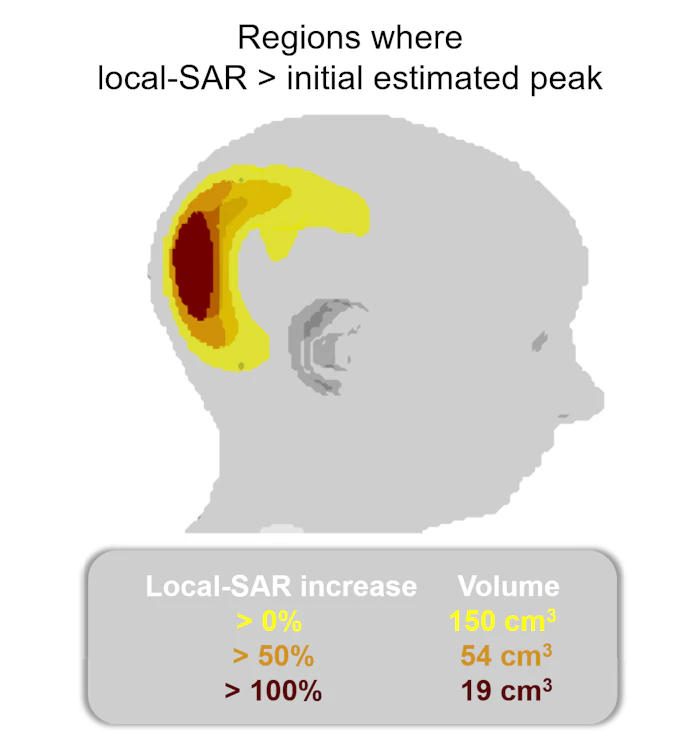
Figure 1: Subject motion during the scan (20 mm rightward) caused peak local SAR to increase more than 3-fold. Results shown for a computational 8-channel parallel-transmit array tuned for 7T.
This is not a problem isolated to cases when the subject cannot remain still. The initial positioning of the subject also has a considerable effect on peak local SAR. When the subject is assumed to be at the centre but is positioned elsewhere, SAR may be underestimated by more than five-fold. Example case: Figure 2. More details: https://doi.org/10.1002/nbm.4876.
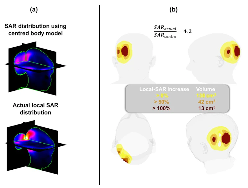
Figure 2: Even with perfect knowledge of the tissue content, a mismatch between assumed and actual subject position can cause substantial SAR underestimation. Results shown for a computational 8-channel parallel-transmit array tuned for 7T.
Preventing motion-related SAR increases require very large safety margins, which reduce imaging performance substantially, making MRI scans much longer. Alternatively, calculations can be adapted to subject motion in real-time. This relies on two conditions:
1- The change in how coil elements interact with tissues is known. This interaction leads to SAR.
2- Changes to the radiofrequency pulse can be performed in real-time.
We have demonstrated that Artificial Neural Networks (here, U-Nets) can be used to estimate how SAR changes reliably, in under 0.2 seconds of computation time. Example case: Figure 3. More detail: https://doi.org/10.1002/mrm.70363.
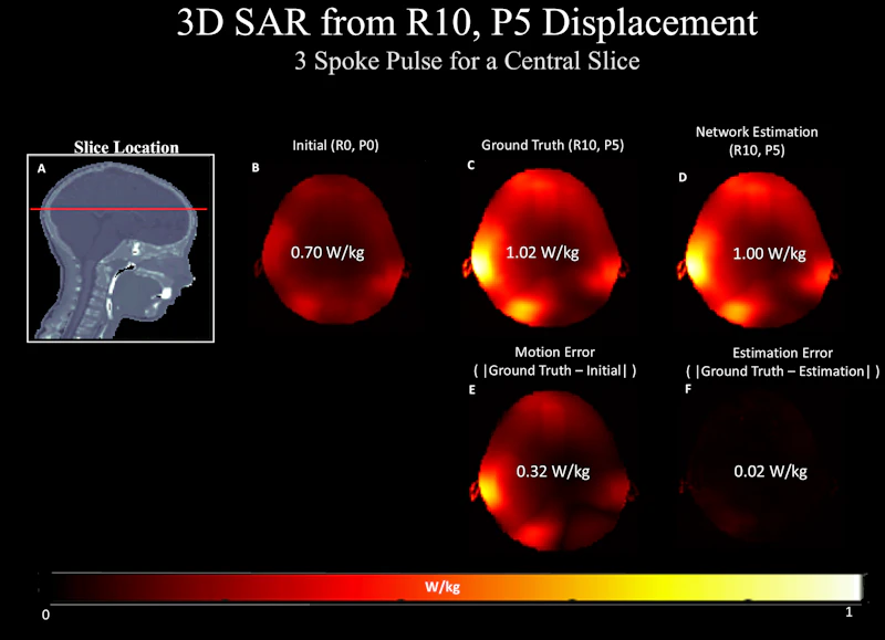
Figure 3: When the subject moves away from their initial position, the peak local SAR is underestimated by 31% (ground-truth: actual peak, initial: estimated using safety model located at centre). U-Nets can recover the actual peak local SAR with less than 2% error. Results shown for a realistic pulse that excites a homogeneous slice, using a computational 8-channel parallel-transmit array tuned for 7T.
Subject motion can cause excitation related data inconsistencies
Subject motion also affects the coil sensitivities inside the tissues. This leads to degradation of excitation homogeneity, creating artificial contrast variations on the image. These variations are unrelated to the tissue, and therefore, reduce diagnostic value. We have also used Artificial Neural Networks (here, cGANs) to estimate the changes in coil sensitivities, which enables improving excitation homogeneity as subject motion happens. Example case: Figure 4. More detail: https://doi.org/10.1002/mrm.29132.
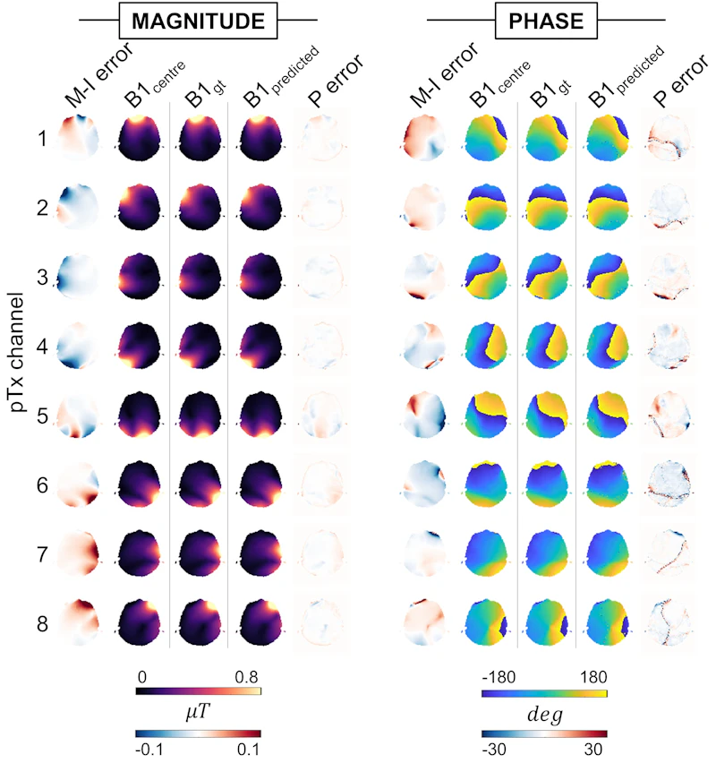
Figure 4: The effect of motion on coil sensitivities is shown. The networks can reliably estimated the effect of motion on coil sensitivities. Results shown for a computational 8-channel parallel-transmit array tuned for 7T.
Shorter scans can yield high quality images when images are processed together
In clinical settings, multiple imaging protocols are used to image a subject. These imaging protocols are adjusted such that each image set is under the influence of a different contrast mechanism (Figure 5). These images provide complementary information, and therefore, maximize diagnostic value.
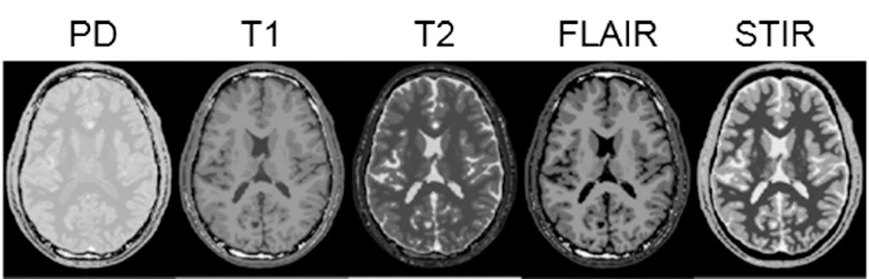
Figure 5: Images acquired under the influence of different contrast mechanisms provide complementary diagnostic information.
To reduce scan time, MRI protocols can be accelerated by acquiring less data. If certain conditions are satisfied, the effect of this data reduction can be compensated for via image processing. When we are processing acquired data, we can process different contrasts together. This allows information sharing, and improves image quality (Figure 7). However, this joint processing may also cause detrimental effects, such as the leakage of features that are unique to an image to the other images (leakage-of-features, Figure 6).
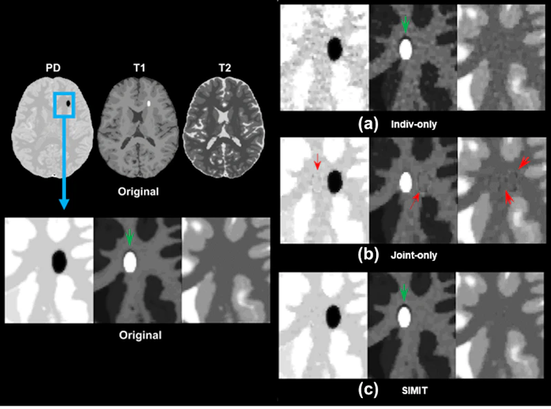
Figure 6: Processing images together (b) improves image quality compared to each image going through nonlinear reconstruction separately (a). However, this leads to leaking of features that are unique to one image to the other images (red arrows). Our proposed reconstruction method suppresses such leakage artefacts and yields artefact-free high-quality images (c). Please note that the image contrast was adjusted to maximize visibility of artefacts.
We proposed an image reconstruction algorithm that processes images both together and separately. Processing images together improves quality while processing images separately ensures that each image is faithful to its data. Therefore, the method yields high-quality images free of leakage-of-features (Figure 6). In-vivo images where the scan was accelerated by 87.5% show that high quality images can be acquired at 12.5% of the duration of a standard protocol (Figure 7).
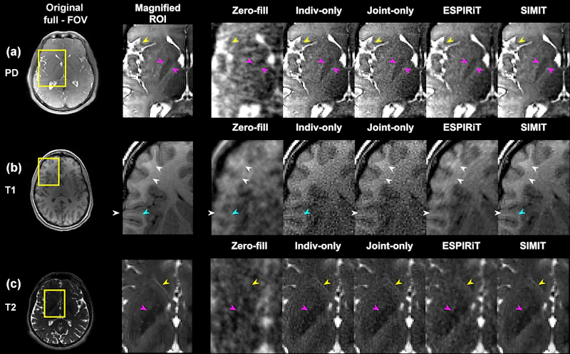
Figure 7: Proton-density weighted, T1-weighted and T2-weighted images were processed together to reconstruct high quality images. All imaging protocols were 87.5% accelerated compared to their standard versions (acceleration factor R=8). The proposed method (SIMIT) showed the Lentiform Nucleus (pink arrows) and the frontal opercular cortex (yellow arrow) more clearly. SIMIT also depicted the gray-matter boundaries in the sulci more clearly in the T1-weighted images.
Blinded and random-order neuroradiologist scores highlight the superior performance of SIMIT in terms of diagnostic value (Figure 8).
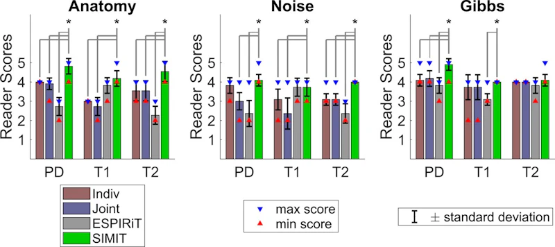
Figure 8: Neuroradiologist scores highlight the improved image reconstruction performance of SIMIT. The neuroradiologist was blinded to method names and images were presented in randomized order.
Funding
|
Wellcome Trust Discovery Award (2025 – 2033) – ~ £4.9M Democratising Neuroimaging Research with MRI Derek Jones, Marco Palombo, Johnes Obungoloch, Emre Kopanoglu, Daniel Alexander, Andrew Webb, Mara Cercignani, Mark Griswold Role: Co-I Status: Active |
|
EPSRC Doctoral Training Partnership – PhD Studentship (2024 – 2028) - £ 85,301 The Beat Goes On Ian Driver, Kevin Murphy, Emre Kopanoglu Role: Co-I PhD Studentship; 2024 – 2028 Status: Active |
|
Cardiff University – PhD Studentship (2024 – 2028) - £ 85,301 Make Yourself (MY-) Magnetic Resonance Imaging (MRI) Scanners: Designing Very Low Cost MRI Scanners to Make Medical Imaging Available in Underfunded Settings Emre Kopanoglu, Derek K. Jones, Mara Cercignani Role: PI PhD Studentship; 2024 – 2028 Status: Active |
|
SPF Research Grant (2023 – 2028) - £ 29,810,359 National facility for ultra-high field (11.7T) human MRI scanning PI: Richard Bowtell. Co-I: Karin Shmueli, Shajan Gunamony, Jurgen Schneider, James Wild, Andrew Peet, Laura Parkes, Christopher Rodgers, Paul Glover, Ian Hall, Emre Kopanoglu, Zoe Kourtzi, Dorothee Auer, Harish Poptani, Penny Gowland, Shaihan Malik, Andrew Blamire, Damian Tyler, Andrew Bagshaw, Neal Bangerter, Geoff Parker, Derek Jones, Susan Francis, Paul Armitage, Jozien Goense, Peter Jezzard, Adam Berrington, Mara Cercignani, Ozlem Ipek, Steven Williams, Karen Mullinger, Rimona Weil, James Rowe, Daniel Alexander, Steven Sourbron, Peter Thelwall, Stuart Clare, Claudia Wheeler-Kingshott, Andrew Peters , Itamar Ronen Role: Co-I Status: Active |
|
BBSRC Mid range equipment Initiative (2023) - £ 860,000 Upgrading our view of Growing Older: Mapping Brain Changes across the Lifespan with Ultra High Field Multi-Spectral MR Mara Cercignani, John Evans, Derek Jones, Emre Kopanoglu, Michael Germuska, Daniel Gallichan, Kevin Murphy, Robert Turner Role: Co-I Status: Active |
|
EPSRC Doctoral Training Partnership – PhD Studentship (2021 – 2025) - £ 81,528 New methods to quantify axonal magnetic properties and myelin integrity using MRI Marco Palombo, Emre Kopanoglu, Robert Turner Role: Co-I PhD Studentship; 2023 – 2027 Status: Active |
|
Cardiff University Neuroscience and Mental Health Innovation Institute – Future Leaders in Neuroscience Research Award (2023) - £ 1,410 Conference travel support Status: Complete |
|
Welsh Government Data Nation Accelerator Award (2022) - £ 8,836 Improving image quality and safety of ultra-high field magnetic resonance imaging using deep learning-based electromagnetic field prediction Emre Kopanoglu, Alix Jean Deeley Plumley, Kevin Murphy Role: PI Status: Complete |
|
EPSRC Doctoral Training Partnership – PhD Studentship (2021 – 2025) - £ 72,404 Using machine learning to ensure safety of patients who cannot remain still during magnetic resonance imaging Emre Kopanoglu, Kevin Murphy Role: PI PhD Studentship; 2021 – 2025 Status: Active |
|
Wellcome Trust Seed for Seed Award (2018) - £ 20,000 Magnetic resonance imaging of moving patients at ultra-high field: motion exacerbates the homogeneity artefacts due to wavelength effects Emre Kopanoglu Role: PI Status: Complete |
|
EPSRC Doctoral Training Partnership – PhD Studentship (2018 – 2022) - £ 71,101 Patient-motion tolerant functional Magnetic Resonance Imaging at the Ultra-high Field Emre Kopanoglu, Kevin Murphy, Richard G. Wise. Role: PI PhD Studentship; 2018 – 2022 Status: Complete |
|
EPSRC Doctoral Training Partnership – PhD Studentship (2018 – 2022) - £ 71,101 Magnetic Resonance Imaging of Moving Patients at Ultra-high Field: Real-time Motion Corrected Parallel-transmit Pulse Design Emre Kopanoglu, Kevin Murphy Role: PI PhD Studentship; 2018 – 2022 Status: Complete |
Research group
Teaching
Qualifications
|
Fellow |
Higher Education Academy, UK |
2023 |
|
Scientific Teaching Fellow |
Yale University, New Haven, CT, USA |
2014 |
Modules taught
PST 518 In-vivo Human Imaging
PS 3214: Neuroimaging in Health and Disease
PS 1018: Research Methods in Psychology
PS 3003: Occupational Placement
PST 510: Neuroimaging Research Project
PST 512: Introduction to Neuroimaging Methods
PST 513: Research Design and Analysis in Neuroimaging
PST 514: Introduction to Statistics and Matlab Programming
PST 515: Neuroimaging Research Proposal
Other Roles
Guest Lecturer, Cardiff University School of Engineering, 2022 – 2024
CUBRIC Health and Wellbeing contact, 2024 –
Deputy Lead for Extenuating Circumstances, Cardiff University School of Psychology, 2022 –
Disability Officer, Cardiff University School of Psychology, 2022 –
Ethics Committee member, Cardiff University School of Psychology, 2022 –
MSc Programme Deputy Lead & Acting Co-Lead, Cardiff University School of Psychology, 2021 – 2022
Biography
Education
- 2012: PhD in Electrical and Electronics Engineering. Bilkent University, Ankara, Turkey. Novel Techniques Regarding Specific Absorption Rate and Field of View Reduction in Magnetic Resonance Imaging
- 2006: BSc in Electrical and Electronics Engineering. Bilkent University, Ankara, Turkey.
Employment
- 2017 – present: Lecturer / Senior Lecturer in Psychology. Cardiff University, Cardiff, UK.
- 2015 – 2017: Senior Research Scientist. Aselsan Research Center, Ankara, Turkey.
- 2012 – 2015: Post-Doctoral Associate. Radiology and Biomedical Imaging. Yale University, New Haven, CT, USA.
- 2006 – 2012: Research and Teaching Assistant. Electrical and Electronics Engineering. Bilkent University, Ankara, Turkey.
- 2006 – 2006: Undergraduate Teaching Assistant. Electrical and Electronics Engineering. Bilkent University, Ankara, Turkey.
Speaking engagements
|
Lecturer |
ISMRT, Special ISMRM-ISMRT Forum, Annual Meeting of the ISMRM & ISMRT ISMRM, Weekend Educational, Annual Meeting of the ISMRM & ISMRT ESMRMB, Lectures on MR, Diffusion MRI and Spectroscopy |
2024
2024 2023 |
Committees and reviewing
Committee Experience
Current
| ISMRM Annual Meeting Programming Committee | Member | 2025 – present |
| ISMRM Sustainability Advisory Committee | Member | 2024 – present |
| ISMRM Turkish Chapter | Board Member | 2023 – present |
| ISMRM MRI Safety Committee | Member | 2020 – present |
| Chair | 2021 – 2025 | |
| ISMRM Web Development Committee | Member | 2021 – 2025 |
Organisation
| ISMRM – RadAid International Masterclass Series on MRI Safety for Low- and Middle-Income Countries | Organising Committee Chair | 2026 |
| ISMRT Future Leaders Program Workshop on MRI Safety in collaboration with ISMRM Safety Committee and Rad-Aid International | Organizing Committee Member | 2025 |
| ESMRMB Lectures on MR, Diffusion MRI and Spectroscopy | Organizing Committee Member | 2023 |
| ISMRM British & Irish Chapter Annual Meeting | Organizing Committee Vice-Chair | 2022 |
Supervisions
Current supervision


Past projects
Previous PhD students
- Alix Plumley
- Luke Watkins
- Bleddyn Owen Woodward
- Katherine Anna Blanter
Contact Details
+44 29225 10256
Cardiff University Brain Research Imaging Centre, Floor 1, Room 1.016, Maindy Road, Cardiff, CF24 4HQ
Research themes
Specialisms
- Simulation, modelling and programming of medical imaging
- Magnetic Resonance Imaging
- Patient safety
- Signal and Image Processing
- Compressed Sensing